
Biopolymers for Surgical Applications


1
Department of Morpho-Functional Sciences II—Pharmacology and Clinical Pharmacology, Faculty of Medicine, Grigore T. Popa University of Medicine and Pharmacy of Iași, 16 University Street, 700115 Iasi, Romania
2
3rd Surgery Clinic, Saint Spiridon University Clinical Emergency Hospital, 700111 Iasi, Romania
3
Leather Engineering Department, Faculty of Engineering, Ege University, Bornova, 35100 Izmir, Turkey
4
Department of Analytical Chemistry, Faculty of Pharmacy, “Grigore T. Popa” University of Medicine and Pharmacy of Iași, 16 University Street, 700115 Iasi, Romania
5
Department of Surgery, Faculty of Medicine, Grigore T. Popa University of Medicine and Pharmacy, 16 Universitatii Street, 700115 Iasi, Romania
*
Author to whom correspondence should be addressed.
Coatings 2022, 12(2), 211; https://doi.org/10.3390/coatings12020211
Submission received: 29 December 2021
/
Revised: 25 January 2022
/
Accepted: 3 February 2022
/
Published: 6 February 2022
(This article belongs to the Special Issue Biocompatible Polymer-Based Materials: Synthesis, Properties and Applications)
Abstract
:Biopolymers have gained significant attention as a class of polymer materials with a wide range of applications, especially in the medical and pharmaceutical field. Due to particular characteristics, such as biocompatibility, biodegradability, non-toxicity, and functionality, they have become promising candidates for various surgical applications, including as bioadhesives, sealants, wound dressings, sutures, drug carriers, coating materials, etc. Recent research shows that further modification of biopolymers by advanced techniques can improve their functionality i.e., antibacterial activity, cell viability, drug-releasing capability, good wet adhesion performance, and good mechanical properties. This mini review aims to provide a brief report on the type of biopolymers and recent developments regarding their use in various surgical applications.
1. Introduction
Rapid advances in medical technology have resulted in the improvement of patient care, compliance, and expectations. Significant developments have been made in the technology of the materials used during surgical operations. Surgical procedures, in particular, involve an instrumented incision made in an operating room and anesthesia and/or respiratory assistance. Surgical materials such as tissue adhesives, sealants, hemostatic agents, wound dressings, absorbent sponges, and suture materials are expected to be non-toxic, biocompatible, and to support cell proliferation for tissue healing. They should have sufficient long-term mechanical and physical properties. Biopolymers from natural sources are macromolecules similar to the extracellular matrix; therefore, they are recognized by the body and are biodegradable. Moreover, these biopolymers do not usually induce acute or chronic immune system responses or toxic effects, which are often induced by synthetic polymers [1,2,3]. Surgical interventions are usually specific and sensitive actions, and the motivation to select appropriate systems to improve patient compliance, health, and comfort is very high. Therefore, biopolymers represent excellent options for surgical materials. With the recent developments in chemistry and material science, the number of studies evaluating the improvement of functionality, physical–mechanical properties, regenerative and adsorption properties, etc., of biopolymers-based surgical materials is constantly growing. For this reason, this mini review aims to highlight the use of natural polymers in surgical applications. The first section of the review provides information on the type and structural particularities of biopolymers used for surgical applications and the second section provides an overview of current studies on the use of biopolymers in various surgical applications.
2. Biopolymers Used for Surgical Applications
Biopolymers are mainly naturally occurring polymers, usually obtained from microorganisms or by extraction from plants. Some biopolymers can also be obtained by chemical synthesis from basic biological systems. Their main advantage compared to synthetic polymers is biocompatibility. The structure of biopolymers is more complex, but it is well-defined. They also have functional properties, and degradability and renewability characteristics [4,5,6,7]. On the other hand, their disadvantages are poor mechanical properties, lower productibility, and dependency on many environmental factors; however, proper functionalization can significantly improve their properties. The most commonly used biopolymers in surgical materials are chitosan, alginate, and hyaluronic acid as polysaccharides; collagen, gelatin, fibroin as proteins; and/or polylactic acid derivatives biocompatible polymers. The next section highlights the general properties of main biopolymers and their roles in surgical applications.
2.1. Chitosan
Chitosan, a semisynthetic material derived from deacetylated chitin (Scheme 1), is very similar to cellulose, but has a rigid crystalline structure. The functional hydroxyl group on the C2 position of the cellulose is replaced by acetylamino group in chitosan, while inter- and intramolecular hydrogen bonding is taken into account for its rigidity. Thus, chitosan is a cationic linear polysaccharide consisting of D-glucosamine (2-amino-2-deoxy-D-glucopyranose) molecules and N-acetyl glucosamine (N-acetyl-2-amino-2-deoxy-D-glucopyranose) molecules variably distributed in the polymer, linked by β-(1,4)-D-glucosamine bonds [8].
The antifungal or antibacterial effects of chitosan are due to the polycationic nature of chitosan in acidic medium (pH < 6). The antimicrobial action of chitosan is thought to be induced by the existing electrostatic charges between chitosan amine (NH3+) groups and negatively charged moieties existing on the cell surfaces. When the deacetylation degree (DD) of chitosan is higher, the amount of positively charged amine groups also increases, which affects antimicrobial activity. Moreover, chitosan also forms a film material with mechanical properties, high permeability to water vapors, and selective permeability to CO2 and O2. However, the high permeability to water vapor limits the use of chitosan, since effective control of moisture transfer is a desirable property in moist mediums. Due to its characteristics of being biodegradable, biocompatible with bioadhesive and antimicrobial effects, and having relatively good mechanical and barrier features, chitosan may be an ideal candidate to create bio-nanocomposites with different practical uses [9,10,11,12].
Recently, chitosan and its derivatives were reported by Feng et al. [13] to play important roles in the first three stages of wound healing, especially hemostasis, inflammation, and proliferation. In the first stage of hemostasis, it helps to stop bleeding by initiating the aggregation of platelets and red blood cells, followed by the second stage called hemostasis, characterized by the inhibition of the dissolution of fibrin. Further, during the inflammation stage, bacteria are removed from the wound. In the final stage, development of granulation tissue contributes to the rapid healing of the skin.
2.2. Alginate
Alginic acid is a polyuronide that is a biopolymer based on polysaccharides extracted from brown algae. Due to its biocompatibility and biodegradability, alginic acid has encountered a wide range of research interests. Due to its unique physical properties, it can be used as biomaterial for medical or pharmaceuticals products. It can also be used in other domains as a rheology modifier for food or printing inks.
Alginic acid has repeating units of α-L-guluronic acid (G) and its C5 epimer β-D-mannuronic acid (M), which are linked by 1,4-glycosidic bonds. The orientation of the carboxyl group (–COOH) on the C5 carbon of the six-membered sugar ring is above the ring plane of the M unit and below the plane of the G unit (Scheme 2).
The alternating sequences of guluronic acid (G-block), mannuronic acid (M-block), or of both (MG-block) form the polymer chain. Viscosity and the ability to form gels are the most important properties of alginate, which are strongly influenced by the block structure and length of the chain [14].
Recent developments in natural polymers used to heal injuries/trauma have highlighted the specific characteristics induced by alginate-based systems. The main requirements for an ideal wound healer should be good adhesion to the injured surface, biocompatibility with the human body, non-antigenic, with enough permeability, biodegradable, elastic but with tensile at break, and easy to be produced.
An ideal wound healer should have bacterial inhibitory activity to prevent infections; in addition, it should improve mechanical properties and be thermally sensitive, regulate the appropriate level of moisture, encapsulate, and transport different types of molecules to the target site.
Barbu et al. [15] reported the contribution of alginates in various complex wound dressings. Specifically, alginates and their derivatives have been shown to have anti-bacterial effects on Gram-positive bacteria (such as Staphylococcaceae or Bacillus cereus) and Gram-negative bacteria (such as Escherichia coli, Pseudomonas aeruginosa, and Acinetobacter spp.), antifungal activity against Candida albicans, and antiviral effect against Herpeviridae, Rhabdoviridae, Flaviviridae, and Togaviridae. Alginates and their derivatives have also been reported to have anti-inflammatory effects, and regenerative and angiogenetic properties. Their immunomodulatory and antioxidant effects have been shown to be due to the release of nitric-oxide (NO), the release of reactive oxygen species (ROS), the release of tumor necrosis factor (TNF-α) and nuclear factor kappa-light-chain-enhancer of activated B cells (NF-KB) from macrophages, and through activation of the mitogen-activated protein kinases (MAPK) signaling pathway. Their hemostatic effects were produced by platelet activation and thrombin clot formation.
2.3. Hyaluronic Acid and its Derivatives
Hyaluronan or hyaluronic acid (HA) a natural polymer belonging to the group of heteropolysaccharides known as glycosaminoglycans (GaGs). GaGs are found mainly in the vitreous humor, joints, skin, and connective tissue in humans. HA or hyaluronan, consisting of repeating units of an uronic acid (β-d-glucuronic acid) and an amino sugar (N-acetyl-d-glucosamine), is linked alternately via β-(1,3)-D-glucosamine bonds and β-(1,4)-D-glucosamine bonds (as shown in Scheme 3).
Each carboxyl group found within HA’s structure ionizes at physiological pH. The anion resulting from ionization forms hydrogen bonds with the water molecules through carboxyl groups of the side chain and planar acetamido groups of this biopolymer. Thus, these bounds stabilize the secondary structure of HA. As the pH value of the wound bed may fluctuate during the healing process, such effects of pH on the properties and effectiveness of HA should be taken into consideration when using HA for wound healing. The pH value of an injured area increases to about 8 after injury, but when the healing process stops, the pH drops to ≈5 [16,17].
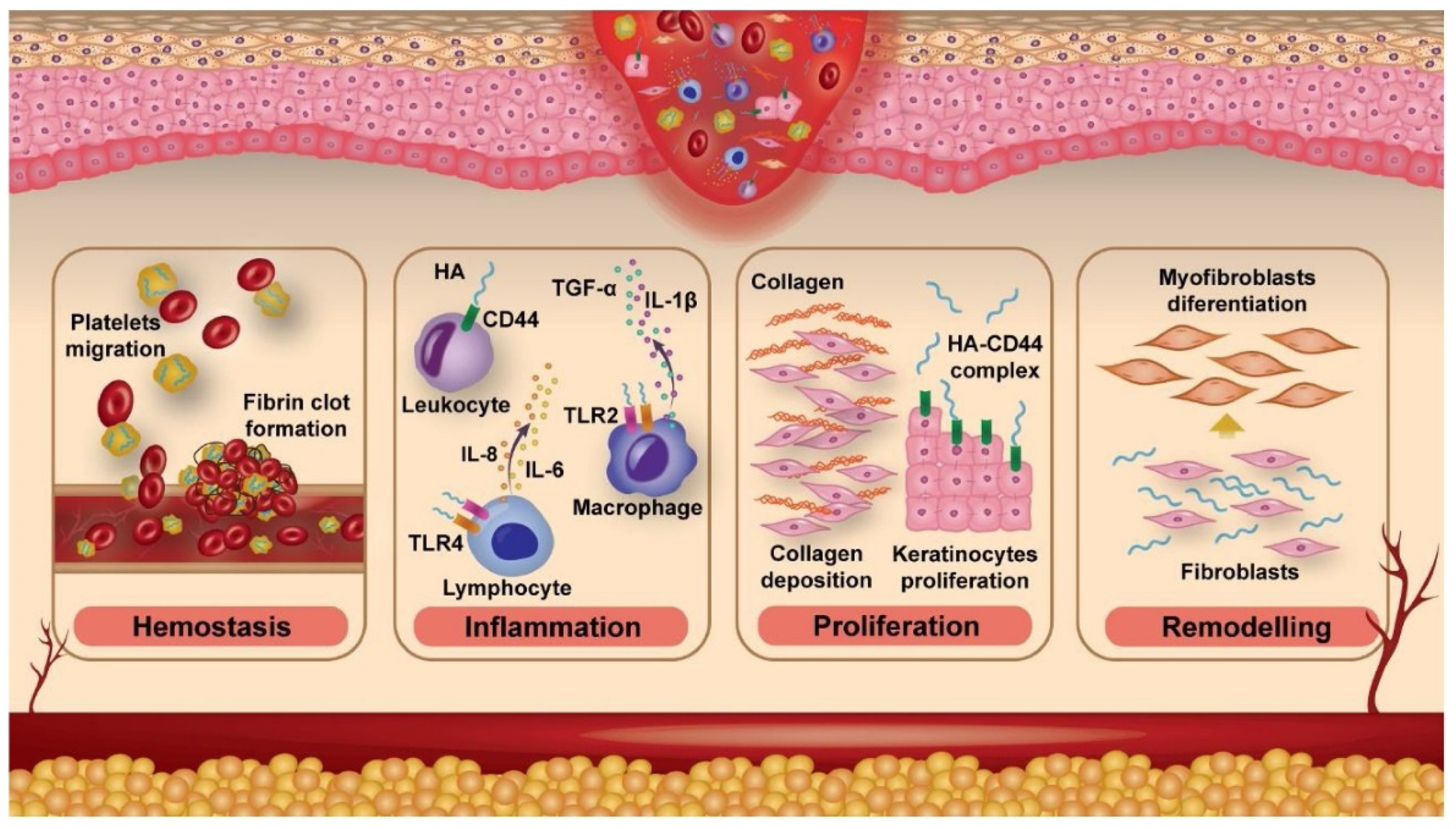
Graca et al. [18] highlighted the roles of HA in the wound-healing process. They found out that hyaluronic acid is involved immediately after the incision or injury occurs, the healing activity starting with the restoration of the skin tissue architecture and stopping the bleeding. Furthermore, a large number of platelets are released due to the presence of high molecular weight of HA, which further promotes fibrinogen deposition and initiation of coagulation cascade. As the main component of edematous fluid, HA is responsible for the recruitment and migration of neutrophil cells to debris and dead cells to eliminate them by phagocytosis, followed by the release of cytokine necrosis factors (i.e., tumor necrosis factor TNF-α, interleukins IL-1β, and IL-8).
In the final stage of the inflammatory phase, lymphocytes and macrophages travel to the site of the lesion to interact with low molecular weight HA fragments (LMW-HA) through Toll-like receptors (TLR) type 2 (TLR2) or type 4 (TLR4), resulting in the release of cytokines such as TNF-α, IL-1β, IL-8, and IL-6. The main roles of HA in the healing process are summarized in Figure 1.
2.4. Collagen
Collagen, as a biopolymer, is the major component of the extracellular matrix (ECM) of the human body. The cell environment of a tissue depends on the detailed molecular structure of collagen and defines their properties, such as differentiation and metabolic states. The interaction between tissue and external stress stimuli depends on the specific mechanical properties provided by self-assembled collagen fibrils and the well-characterized protein binding sites at the molecular level for interaction with cells [19].
During the wound-healing process, collagen molecules are synthesized by fibroblast cells with different morphologies and their type, structure, mass and fiber network are modified during healing. For example, at the beginning of the wound healing, first of all, Collagen III is synthesized and then it is transformed into Collagen I as the most abundant skin type. Tissue formation involves the initial deposition of collagen, which is enhanced by covalent crosslinking via lisle oxidase enzyme. This maturation changes collagen into more stable structures that significantly increase tensile strength. The process of changing collagen in the repaired tissue usually continues for several months after the wound has closed.
The most common types of collagen are type I, III, and V, which are found in the skin. The other collagens, type IV and XVIII, as non-fibrillary types, are usually located at the lower membrane in the skin.
In skin and wounds, collagen contributes to the inflammatory phases of wound healing, such as hemostasis and inflammation. As a structural component of ECM, collagen also plays an important role in its remodeling, providing important support for vascular development in angiogenesis, and thus in the healing process.
Mathew–Steiner et al. [20] reported that collagen administered as augmentation therapy for injury recovery could induce healing, acting as a trap for the matrix metalloproteinases (MMPs) and other enzymes at the injury site, thereby reducing inflammatory factors and supporting recovery phases (as summarized in Figure 2).
2.5. Silk Fibroin
Silk fibroin, produced by the Bombyx mori silkworm, has been considered a high quality fiber for centuries. In addition to textiles, silk fibroin was proposed many decades ago for use in medicine as a surgical suture material and later developed for a variety of new applications in medicine [21].
Silk fibroin (SF) is reported to be composed of a light (L) chain polypeptide and a heavy (H) chain polypeptide, bound by a disulfide bond at the C-terminal of the chain, forming an H–L complex. The H-chain forms β-sheet crystallites, which are the major structural components involved in mechanical role, and the L-chain has a much smaller mechanical role due to its smaller size compared to the H-chain. In terms of amino acid composition, the H-chain contains mainly the three simplest amino acids: glycine (G) (43%–46%), alanine (A) (25%–30%), and serine (S) (12%). Tyrosine (Y), the largest amino acid with a polar side chain, is about 5%. Other most common amino acids in the H-chain are valine (V) (2%), and, to much lesser proportions, aspartic acid (D), phenylalanine (F), glutamic acid (E), threonine (T), isoleucine (I), leucine (L), proline (P), arginine (R), lysine (K), and histidine (H).
Over time, SF has always been known as a fiber-based material with excellent mechanical properties, high tensile strength, and breaking strength. SF has been used as a suture material for over 2000 years due to its advantages, such as very good biocompatibility, controlled biodegradation, and low toxic effects.
It is also known that SF is involved in various stages of wound healing, starting with the stimulation of the proliferation, growth, and migration of various cells.
Tanaka et al. [22] reported 1.5 mm diameter double-raschel knitted elastin (EL)–SF vascular graft for better cell proliferation.
In the case of dermal injuries generated on the backbone of test rabbits, silk fibroin hydrogels showed better recovery than conventional wound dressings, the epidermal reconstruction of which was confirmed by histological analysis. Pollini et al. [23] also reported that wound size was reduced to 30% after 10 days and to 11% after 15 days of treatment with silk fibroin material, while control samples resulted in reduction of 52% and 49%, respectively.
A beneficial effect on the healing of hypertrophic scars has been reported when silk fibroin is applied on the surface of the skin. These scars are associated with high ECM protein deposition in fibroblasts, as well as permanent inflammation and fibrosis. When applied, the fibroin hydrogel changed the color of the scar to white and decreased its thickness [24]. Silk fibroin enhances mechanical properties and supports physical stimulation for endothelial cells differentiation and angiogenesis without additional growth factors [25]. These effects of silk scaffolding on cell adhesion, migration, and differentiation were found to be correlated with the structure of the scale.
3. Applications of Biopolymers for Surgical Needs
Due to the biocompatibility, biodegradability, functionality, and non-toxicity nature of biopolymers, they are highly preferred for medical applications, including surgery. Applications that may be mentioned for surgical needs are usually focused on adhesives, sealants, hemostats, sutures, and surgical instruments. Biopolymers are usually applied to current surgical materials to increase their functionality, such as antibacterial activity, cell viability, adhesion, mechanical properties, etc. Some examples of the use of different biopolymers in surgical applications are summarized in Table 1. More details on the various uses of biopolymers in surgery are provided in the following sections.
3.1. Adhesives and Sealants
The sealing of instant ruptured/incised tissues during surgery is essential for patients’ safety. Classical techniques such as sutures and staples used to connect the incisions offer the disadvantages of long duration, risk of damage to tissues and infections, and low water resistance (thus, they can lead to bleeding) [38,39,40]. Therefore, surgical sealants have gained significant interest as alternatives for wound closures due to ease of application, shorter delivery time, improved sealing, and less pain [41,42]. Different types of surgical adhesives obtained from synthetic or natural-based polymers or combinations can be used for closing, restoring tissue continuity, or attaching devices to tissues.
On the other hand, it is very difficult to ensure significant adhesion to soft tissues together with the properties of being nontoxic and provide non-tissue damage or side-effects. Another challenge for the sealants is to provide acceptable adhesion strength in humid and dynamic environments, especially when blood is present. Currently available sealing materials are mostly based on cyanoacrylates and fibrin-based materials. Cyanoacrylates are fast-reacting sealants that solidify rapidly after contact with blood; however, they become too rigid, are not compatible with soft tissue, and have high toxicity, limiting their use [43,44]. A fibrin-based sealant has been reported as being biocompatible and nontoxic; however, they lack sufficient adhesion to bleeding sites and low mechanical resistance at the environment. Therefore, there is a high demand for biocompatible, nontoxic, soft sealants, with high moisture adhesion.
There are many studies to improve the performance of the surgical sealants based on biopolymers. While studying the source of shellfish that release catechol-rich proteins to glue themselves on rocks in a dynamic water environment, Bai et al. [38] prepared a silk-based sealant (SFT) by the addition of tannic acid (TA) to silk fibroin. The existence of high phenolic moieties from TA helped transform a random structure on fibroin into b-sheet form and lead to hierarchical assembly of nanofibrillar structures. The final sealant had a higher mechanical toughness (123.1 kJ·m−3), improved adhesion in a wet environment (134.1 kPa), and induce rapid hemostasis (in less than 30 s). In vivo experimental models in rats showed rapid closure of severely injured tissues, with loss of blood in humid and dynamic environments. They also mentioned that the sealants had good biocompatibility, ability to degrade rapidly, and antibacterial properties [38].
In a study using other biopolymers as starting materials, Gasek et al. [45] prepared two sealants composed of alginate methacrylate and gelatin methacryloyl, which were further functionalized by chemical modification through conjugation with dopamine hydrochloride to be used for pleural and tracheal lung injuries. In the experiments, methacrylic anhydride was added drop by drop to sodium alginate and gelled separately under basic conditions. After completion of the reaction, alginate methacrylater (ALG-MA) and gelatin methacryloyl (GEL-MA) solutions were dialyzed and lyophilized. Both materials were subjected to a reaction schema mediated by carbodiimide-mediated reaction under the protection of nitro gas, resulting in two dopamine-conjugated compounds: dopamine-conjugated alginate methacrylate (ALG-MA-DA) and dopamine-conjugated gelatin methacryloyl (GEL-MA-DA). Methacrylation of biopolymer materials resulted in a functional group able to be activated by light, depending on the existence of a photo-initiator, to a covalent crosslinking by free radical polymerization. As example, Eosin Y used visible or monospectral light, which is an advantage over cross-linking with the UV light spectrum [46].
The conjugation with dopamine, similar to most important adhesive functional groups in mussel-foot proteins, has been used to increase adherence in moist/wet conditions [47,48,49,50]. Therefore, both compounds were crosslinked into pharmaceutical dosage forms such as patches with a pre-formed hydrogel or as hydrogels placed at the lesion site. Testing of the materials showed that both sealants had sufficient adhesion in wet conditions, tensile strength, burst pressure, and elasticity without important toxicity to the cells, as shown in in vitro experiments. Moreover, no air leakage was observed after the ex-vivo administration to experimentally-induced lung lesions in rats and in pig and tracheal lesions in rats (as shown in Figure 3). The recovery time of the lesions was noticed to be up to a month for the lung lesions from rat or pig models and up to two weeks for the tracheal lesions from rat models (Figure 4). As a major outcome, the alginate-based sealant performed better for patches with a pre-formed hydrogel, whereas the gelatin-based sealants were more effective than the in situ application of hydrogels to be formed at the lesion site [45].
A recent study by Rajabi et al. (2020) [51] reported the use of an injectable hydrogel for surgical sealing based on thiolated gelatin (Gel-SH), gelatin methacrylate (GelMA), and polydopamine functionalized Laponit (PD-LAP). Initially, they synthesized thiolated gelatin, where carboxyl groups of gelatin were first aminated with ethylenediamine, followed by a reaction with 2-iminothilane to obtain thiol end functional groups. Then, they prepared gelatin methacrylate by dropping methacrylic anhydride into gelatin solution in a controlled environment. In the next step, polydopamine-modified Laponit was obtained by adding a solution of dopamine in the Laponit nanoplates suspension and maintaining it at room temperature for 5 h to allow dopamine to intercalate and oxidize. The final nanocomposite hydrogels were obtained by mixing the first 5 wt.% GelMA solution with solutions of different concentrations of PD-LAP (0.5, 1 and 2 wt.%). A solution with a concentration of 1.75 wt.% of Gel-SH was mixed with GelMA/PD-LAP and the resulting hydrogel solutions were formed by crosslinking in visible-light using photoinitiators and an N-vinylcaprolactam monomer. They analyzed the differences in physical properties among the newly obtained nanocomposite hydrogels with various concentrations of PD-LAP: pore size, mechanical properties, swelling behavior, and rate of degradation. They found that the Gel-SH/GelMA hydrogel obtained by the incorporating 1 wt.% PD-LAP presented a 4.4-fold increase in tensile strength, a 9.3-fold increase in toughness, and a 2-fold increase in elongation, and used the cyclic compressive test to evaluate its dynamic properties. The main result found for Gel-SH/GelMA hydrogel was that PD-LAP nanoparticles determined an improvement in adhesive strength to the tissues, an increased ability to coagulate blood, and good biocompatibility. In addition, the nanocomposite hydrogel significantly reduced the blood clotting time (1.5 ± 0.5 min), compared to the control products (HelistatTM® and AviteneTM® hemostatic surgical sealants), which was finally demonstrated to be a promising hemostatic hydrogel [51].
Another natural biopolymer for parenteral administration based on hydrogel adhesives/sealants with the ability of thermo-reversible adhesion on the surface of the tissues was demonstrated by Zhou et al. (2021) [29]. Hydrogels consist of chondroitin sulfate (CS) mixed with gelatin (Gel). First, CS was mixed with sodium periodate aqueous solution to obtain functional aldehyde CS. Next, by forming a dynamic Schiff base bond, a hydrogel adhesive was produced that could be easily injected from different ratios of CS-Gel and CS aldehyde mixtures in the presence of borax. The results showed that adjusting the CS gel concentrations and the CS oxidation degree of the hydrogel adhesives influence the mechanical properties, the adhesion strength, and breaking pressure. Due to the reversible nature of Schiff base interactions, injectable hydrogels have been shown to be self-healable and remodelable, allowing for simple surgical use during interventional procedures, their administration being with syringes or other delivery systems. On the other hand, tissue adhesion at normal physiological temperature (37 °C) was strong, while the tissues adhesion at low temperature (20 °C) was low due to of the Shiff base bonds and the intrinsic thermal sensitivity of gelatin-based hydrogels. They also demonstrated that the adhesives were effectively used in treating fatal bleeding liver wounds and immediately sealing damaged uterine tissue without the need for sutures or staples [29]. As can be seen from the examples summarized above, studies on biocompatible adhesives and sealants are fast growing, providing enhanced properties and functionality to surgical materials. However, the topic seems to be open for further developments, especially for high wet adhesion and proper mechanical performance with the combination of the ability to regenerate new cells or tissues.
3.2. Sutures
Wound closure is defined as being the final step of surgical intervention [52]. Two major types of wound closure have been identified: primary and secondary. The approach is different at the end of surgery intervention: in primary closure, the skin wound is closed with sutures, whereas in secondary closure, the wound remains open and healing take place through granulation and contraction processes.
The main material used for would closures during surgeries was suture material. These are monofilament and multifilaments implants that join the tissue edges together or compress blood vessels to stop bleeding until the natural healing process provides adequate healing [53]. A large number of materials for sutures are available, and the surgeon can make a choice from materials with different properties to determine the one best suited to their needs.
When choosing a suture material suitable for closing and healing the wound, the following parameters must be taken into account: suture resistance, tissue-retention capacity, absorbency, risk of infection, and inflammatory response induced by the suture material. Other factors to be taken into consideration include: the type of incision, the technique of suture, and aspect of the wound site.
Suture materials usually can be divided into two groups of absorbable or nonabsorbable sutures, which are made of natural or synthetic materials, respectively [54]. Non-absorbable sutures are usually used to close external wounds and are normally removed by a secondary operation, while absorbable sutures can be used for both internal and external wound closure.
The oldest natural and degradable suture material is known as catgut [55]. A few years after its introduction, catgut sutures became unquestionably associated with hypersensitivity reactions and an unusually high frequency of infections; therefore, a lot of surgeons refuse to use them and blame the use of catgut sutures for problems [56]. Consequently, there is an increasing demand for the use of biocompatible and biodegradable suture materials based on natural polymers.
Silk is one of the most commonly used natural non-absorbable suture material due to its various advantages, such as excellent tissue compatibility, high suppleness, easy handling, and strength of knot. However, a major difficulty in using silk is its low resistance to microbial attacks and mechanical properties that can adversely affect wound healing.
Recently, it has been reported that various functional suture materials based on biopolymers extend the tissue’s ability to close wounds. Shubbaa et al. (2019) [57] prepared natural silk fibers coated with ZnO nanoparticles to improve their antibacterial and mechanical properties as a suture material. They synthesized ZnO nanoparticles using natural honey solutions as an agent for reducing and stabilizing Zinc acetate under hydrothermal conditions. The degummed natural silk fibers consist of tufts of fibers with 40–45 filaments/tuft that have been mixed with the ZnO nanoparticle solution under optimized conditions related to time (optimum value of 1 h), pH (optimum pH value of 7.7), and temperature (optimum value of 40 °C). The antimicrobial activity and mechanical properties of the suture material were analyzed. The important antibacterial effect of fibers coated with ZnO NPs on Staphylococcus aureus (S. aureus) was found for up to six days. Powder X-ray diffraction (PXRD), scanning electron microscopy (SEM), and energy dispersive X-ray analysis (EDS) analyses show that the mechanical strength of coated fibers is superior to that of uncoated fibers because the small ZnO nanoparticles crosslink the two brins of a bave. The authors concluded that incorporating ZnO NPs on deguminated silk fibers improves the mechanical strength, thermal stability, and antibacterial effects of natural silk fibers to be used as suture material.
In another study to increase the antimicrobial effects of silk-based suture materials, Franco et al. (2019) [58] used recombinant DNA technology. Their purpose was to obtain new materials with functionalization of spider silk chimeric proteins with antibacterial peptides in order to use them as a coating on commercially silk surgical sutures for the prevention or reduction of postoperative infections on a surgical site. The pET30 vector carrying the 6mer recombinant spider silk protein was inserted with the DNA encoding the antimicrobial peptide HNP to obtain the 6mer-HNP1 biotechnology-derived spider silk protein. The 6mer-HNP1 and 6mer proteins were produced using bioengineering in Escherichia coli (E.coli) and these proteins were subsequently further purified. Next, a commercially available silk suture from the control group (6mer) was coated with spider silk protein and silk suture (Perma-Hand®) of the treatment group (6mer-HNP1) was coated with spider silk protein containing antimicrobial peptides. In vitro studies on human fetal lung fibroblasts (MRC5) have shown maintenance of cell viability in contact with coated sutures and blood compatibility after contact of coated sutures with red blood cells. In addition, the coating significantly suppressed biofilm adhesion and formation and the sutures coated with 6mer-HNP1 had a 1.5 log decrease in Methicillin-Resistant S. aureus (MRSA), compared to the uncoated Perma-Hand® suture, and a 2 log decrease of E. coli, compared to the uncoated Perma-Hand® suture. Bioengineered spider silk protein did not influence the mechanical properties of Perma-Hand® sutures. Using this method, a new class of drug-free sutures was developed to reduce post-implantation infections.
Kalita et al. (2017) [59] studied a new biocompatible suture biomaterial from Eri silk waste to avoid infection at the surgical site. Silk waste fibers were wrapped around the suture material using a 5-loop technique and an Aloe-Vera hydrogel and acacia gum (PRWSc) with a cocktail of antibacterial active ingredients (amoxicillin and amphotericin B, 9:1) and growth factors (nerve growth factor NGF, 250 ng·mL−1 and epithelial growth factor EGF, 1 ng·mL−1). Further, PRWS was immersed in the coating solution and dried at 37 °C for 24 h (PRWSc). The prepared suture showed antibacterial effects against S. aureus and E. coli, and antifungal effects against C. albicans and was able to effectively prevent bacterial adhesion (Figure 5). The hydrogel made possible low and sustained release of the drug, which was achieved by diffusion of the drug in aqueous medium facilitated by the hydrogel. The hydrogel system has unique properties, such as stimulating wound healing, anti-scar, and antimicrobial effects (Figure 6).
Drug-release sutures have been reported by Deng et al. [60]. They used hot melt extrusion technology to produce sutures filled with diclofenac potassium (DP) based on a mixture of PEG/poly(εcaprolactone) (PCL)/chitosan keratin. For this purpose, keratin and chitosan were first mixed together at a ratio of 50:50 w/w. Pre-weighed fibers of PCL/PEG/chitosan-keratin were used in ratios of 80/19/1, 80/18/2, and 80/16/4 w/w along with DP at ratios of 95/5, 85/15, and 70/30 w/w were prepared by melt mixing in a hot-melt extruder at 63 ± 1 °C for 5–20 min. They evaluated the physical, thermal, mechanical, and drug-release characteristics of the reported sutures mechanical properties, as well as drug release properties. They also evaluated the performance of drug-loaded sutures when in contact with the human keratinocyte cell line HaCat. In addition, the tensile properties of the sutures material were considered to be significantly affected by PEG, chitosan, and keratin additives. The optimal formulation of tensile strength was obtained at a ratio of 80/19/1 for PCL/PEG/chitosan-keratin, respectively. Various combinations of PEG/PCL/chitosan/keratin mixtures achieved rapid and sustained drug release rates. The complex of PCL/PEG/chitosan-keratin with 30 wt.% of diclofenac potassium also showed high cell viability and wound healing rates during in vitro cytotoxicity testing.
Many examples of functional sutures can be found with the transport of different drugs [61,62,63,64], bioactive agents [65], growth factors [66], or having other properties such as being electroactive [67], radiopaque [68], etc. We can see that technological progress extended the applicability and effectiveness of surgical sutures. Important advances in this field of research can be explained by the technological discoveries in materials science. Natural polymers play an important role in improving their properties (both physical and mechanical), biocompatibility, biodegradability, and increasing its functionality as a carrier for drugs, stem cells, proteins, peptides, antibodies, DNA, nanoparticles, etc. in the desired position, thereby improving the therapeutic effect of sutures.
3.3. Coatings
Surface changes of biomaterials are important for improving the biocompatibility of surgical implant materials and devices [69,70]. Biodegradable or biosource polymers can be used to improve surface characteristics, such as biocompatibility, antimicrobial effects, mechanical properties of stainless steel surgical materials, meshes, sutures, etc. [71]. In one attempt, Aydemir et al. [72] tried to produce a degradable coating with antimicrobial effects for implants or medical devices made of stainless steel considering their disadvantage of corrosion resistance in simulated physiological media, the absence of the osteointegration process, and the lack of antibacterial action. Two biopolymer solutions (chitosan and gelatin) and silica–gentamicin nanoparticles were proposed as elements of the developed coatings. First, they prepared solutions of chitosan (33 wt.%) and gelatin (67 wt.%). Second, they used a modified Stöber process to prepare nanoparticles with silica-gentamicin (SiGe NPs) [73]. The final coating composition was made after a mixture of 2 g/L of SiGe NPs with the biopolymer solution. The coatings were deposited by electrophoresis on surgical grade stainless steel substrates using direct current with an EX735M Multi-Mode PSU 75 V/150 V 300 W power supply (Thurlby Thandar Instruments Ld.). Microscopic examination was used to characterize the coating surface, and in vitro performances were studied after immersion in different simulated physiological solutions (phosphate-buffered saline, simulated body fluid, cell culture medium). The authors evaluated the degradation of the coating surface, the release of antibiotics, the attachments of the cells (ST-2 stromal cells), and the antibacterial effects against E. coli and S. aureus. They found that the coatings were distributed uniformly and homogeneously covered the substrate surface. Damage to the chitosan–gelatin coatings was observed even after one day (Figure 7) and it was almost much smoother after 21 days due to the release and degradation of SiGe NPs. The release of gentamicin resulted in antibacterial effects after 24 h, as shown in Figure 8, but cell proliferation was not inhibited in a seven-day culture.
Perkins et al. [74] prepared a biodegradable polymer of polyester urethane urea (PEUU) mixed with an antiproliferative agent (taxol) to create a coating, which was then deposited on a titanium substrate using a custom direct-write inkjet technique to create a multilayer thin film. The direct-write inkjet technology deposited multimaterial coatings on three-dimensional (3D) implant devices for orthopedic and vascular applications (pins, screws, stents, etc.). They found that concentrations of Taxol between 5% and 10% w/w of the PEUU load provided the best anti-proliferative effects. Films with less than 10 coating layers had a lower capacity of integration and adhesion effects with the Ti alloy substrate. On the other hand, films with more than 25 coating layers had the capacity of longer release rates on each drug load. In vitro studies using acute blood contact showed that Taxol-loaded PEUU coatings resulted in important reduction of platelet deposition, compared to the unloaded PEUU surface. An important significant decrease in cell proliferation for Taxol-releasing samples was noticed using proliferation assessment of rat smooth muscle cells.
To increase the functionality of surgical steel biomaterials, Gawad et al. [75] reported the preparation of coatings made of nanoparticles of chitosan (CSNPs) and nanoparticles of cobalt (CoNPs). These nanocomposite coatings were deposited on 316L SS alloy in Hank’s solution containing 1 × 10⁻³ M calcium hydrogen phosphate (a new drug used to facilitate bone healing) at pH 7.4 and temperature 37 °C. They found that the newly developed coatings reduced the evolution of hydrogen, the corrosion rate, and increased the biocompatibility and durability of the tested implant. Muro-Fraguas et al. [76] prepared acrylic acid coatings with various numbers of passes to decrease the adhesion of microbes and the contamination of PLA 3D printed surgical tools. The coatings were obtained by deposition, using plasma-polymerization, on the surface of 3D printed PLA Petri dishes, reducing the biofilm production of multidrug-resistant strains (P. aeruginosa, S. aureus). The authors used strains of P. aeruginosa and S. aureus for analysis (six antimicrobial-resistant clinical samples and two susceptible control). They claimed that the polyacrylic acid coatings provided the surface with a higher hydrophilicity, resulting in the formation of a hydrated layer, the thickness of which depends on the roughness of the surface. They showed that polyacrylic acid coatings were more effective with fewer plasma passages, and a relative biofilm reduction of up to 50%.
Due to its physical properties and biocompatibility, polypropylene (PP) is frequently used as a material to produce meshes, especially for urinary incontinence, treatment of hernia, and pelvic organ prolapse [77]. However, PP mesh transplantation may also have adverse effects, such as pain, rejection as a foreign body, adhesion to organs, and infection [78]. Therefore, there are studies to improve the surface properties of PP meshes to reduce their adverse effects by using biopolymer-based coatings. For this purpose, Houshyar et al. 2021 [79] described a two-step functionalization of a PP network using a dopamine-mediated chloro(triphenylphosphine)gold(I)/nanodiamond coating. First, polydopamine (PDA) was polymerized on the conventional polypropylene (PP) mesh surface and then combined with the antimicrobial chloro(triphenylphosphine)gold(I) (Au) and nanodiamond (ND). They effectively adsorb antibiotics with the aim of destroying bacteria in the surrounding tissues by improving overall hydrophilicity and roughness, due to gold compounds, which gives the coating a unique surface with antibacterial activity. The presence of gold compounds on the surface of the mesh improves its contrast characteristics and allows surgical applications to make it easier to monitor the location of the PP mesh after implantation into the body and to detect possible cracks.
Recently, Marinaro et al. [80] described a method that shows the applicability of human menstrual blood-derived mesenchymal stromal cells (MenSCs) on polypropylene meshes. For this purpose, they first performed different plasma treatments for the functionalization of PP surgical mesh surfaces to improve their hydrophilicity and adhesive for fibrin coating. Second, they tested different fibrin coated for the meshes. Subsequently, the properties of cell adhesion, immunomodulatory capacity, and cell viability were tested. They found an optimal condition using fibrinogen (5 mg/mL) and thrombin (5.5 IU/mL) in terms of viability and adhesion. In addition, fibrin-coated meshes reduced the proliferation of CD4+ and CD8+ T, compared to in vitro stimulated lymphocytes. The authors concluded that fibrin-coated PP meshes can support local administration of stromal cells and reduce the response to inflammation after implantation of surgery mesh.
4. Conclusions and Future Perspectives
The present mini-review reported biopolymers used in surgical applications, emphasizing the preparative procedures, modification, and consequent properties of biopolymer-based surgical materials.
The term biomaterial/biopolymer lies on the borderline between two domains: polymer chemistry and medicine, especially surgery. The present mini review aimed to combine chemical science with medicine, to help scientists select proper materials for a particular application, especially considering the characteristics of biomaterials used during and after surgery.
Most conventional surgical materials are still based on synthetic materials, including nylon, polypropylene, polyacrylate, polybutester, etc. Nowadays, new trends in materials for surgery are focused on natural resource materials with high biocompatibility, non-toxic features, and specific properties aimed at the targeted site. Polymeric systems were used for simple sutures in the past, but now their role has become more important because of the need to heal surgical lesions or wounds, refill the damaged surface by activating the cell proliferation, protect damaged tissues and wounds from bacterial contamination and inhibit the infections, stop haemoragia and reabsorb moisture from the wound. Sensitivity to external media seems to be of increasing importance as scientists can adjust the properties of polymer systems by temperature, pH, or other external reactions. Considering the possibility of changes in biopolymers that confer specific properties, such as sensitivity to external stimuli, gelling ability, susceptibility to chemical modifications, in order to create new materials and with new architecture, they seem to be the future of many surgical materials with multifunctions. On the other hand, a large number of articles in this area have proved that the developments to date are still limited and the potential of these new materials is huge.
Author Contributions
Conceptualization, T.B. and O.Y.; writing—original draft preparation, C.M.G.; writing—review and editing, N.B. and R.D. visualization, C.M.G. and O.Y.; supervision. All authors have read and agreed to the published version of the manuscript.
Funding
This research received no external funding.
Institutional Review Board Statement
Not applicable.
Informed Consent Statement
Not applicable.
Data Availability Statement
Not applicable.
Conflicts of Interest
The authors declare no conflict of interest.
References
- Park, S.B.; Lih, E.; Park, K.S.; Joung, Y.K.; Han, D.K. Biopolymer-based functional composites for medical applications. Progr. Polym. Sci. 2017, 68, 77–105. [Google Scholar] [CrossRef]
- Mano, J.F.; Silva, G.A.; Azevedo, H.S.; Malafaya, P.B.; Souse, R.A.; Silva, S.S.; Boesel, L.F.; Oliveira, J.M.; Santos, T.C.; Marques, A.P.; et al. Natural origin biodegradable systems in tissue engineering and regenerative medicine: Present status and some moving trends. J. R. Soc. Interface 2007, 4, 999–1030. [Google Scholar] [CrossRef] [Green Version]
- Dhandayuthapani, B.; Yoshida, Y.; Maekawa, T.; Kumar, D.S. Polymeric scaffolds in tissue engineering application: A review. Int. J. Polym. Sci. 2011, 290602, 1–19. [Google Scholar] [CrossRef]
- Cziple, F.A.; Antonio, J.; Marques, V. Biopolymers Versus Synthetic Polymers. Anul 2008, 1, 125–132. [Google Scholar]
- Augustine, R.; Rajakumari, R.; Mozeti, M.; George, A. Biopolymers for Health, Food, and Cosmetic Applications. In Handbook of Biopolymer-Based Materials: From Blends and Composites to Gels and Complex Networks, 1st ed.; Thomas, S., Durand, D., Chassenieux, C., Jyotishkumar, P., Eds.; Wiley-VCH: Hoboken, NJ, USA, 2013; pp. 801–851. [Google Scholar]
- Niaounakis, M. Medical, Dental, and Pharmaceutical Applications. In Biopolymers: Applications and Trends; Niaounakis, M., Ed.; Elsevier: Amsterdam, The Netherlands, 2015; pp. 291–405. [Google Scholar]
- Rebelo, R.; Fernandes, M.; Fangueiro, R. Biopolymers in Medical Implants: A Brief Review. Procedia Eng. 2017, 200, 236–243. [Google Scholar] [CrossRef]
- Cheaburu Yilmaz, C.N.; Tuncay-Tanriverdi, S.; Ozer, O.; Vasile, C. Polysaccharide containing gels for pharmaceutical applications. In Polymer Gels: Science and Fundamentals, Kumar Thakur, V.K., Thakur, M.K., Eds.; Springer: Berlin/Heidelberg, Germany, 2018; pp. 231–278. [Google Scholar]
- Ciobanu, C.S.; Iconaru, S.L.; Predoi, D.; Trușcă, R.-D.; Prodan, A.M.; Groza, A.; Chifiriuc, M.C.; Beuran, M. Fabrication of Novel Chitosan–Hydroxyapatite Nanostructured Thin Films for Biomedical Applications. Coatings 2021, 11, 1561. [Google Scholar] [CrossRef]
- Akhtar, M.A.; Hadzhieva, Z.; Dlouhý, I.; Boccaccini, A.R. Electrophoretic Deposition and Characterization of Functional Coatings Based on an Antibacterial Gallium (III)-Chitosan Complex. Coatings 2020, 10, 483. [Google Scholar] [CrossRef]
- Gull, N.; Khan, S.M.; Khalid, S.; Zia, S.; Islam, A.; Sabir, A.; Sultan, M.; Hussain, F.; Khan, R.U.; Butt, M.T.Z. Designing of biocompatible and biodegradable chitosan based crosslinked hydrogel for in vitro release of encapsulated povidone-iodine: A clinical translation. Int. J. Biol. Macromol. 2020, 164, 4370–4380. [Google Scholar] [CrossRef]
- Gull, N.; Khan, S.M.; Butt, O.M.; Islam, A.; Shah, A.; Jabeen, S.; Khan, S.U.; Khan, A.; Khan, R.U.; Butt, M.T.Z. Inflammation targeted chitosan-based hydrogel for controlled release of diclofenac sodium. J. Biol. Macromol. 2020, 162, 175–187. [Google Scholar] [CrossRef]
- Feng, P.; Luo, Y.; Ke, C.; Qiu, H.; Wang, W.; Zhu, Y.; Hou, R.; Xu, L.; Wu, S. Chitosan-Based Functional Materials for Skin Wound Repair: Mechanisms and Applications. Front. Bioeng. Biotechnol. 2021, 9, 650598. [Google Scholar] [CrossRef]
- Cheaburu Yilmaz, C.N.; Vasile, C.; Ciocoiu, O.N.; Staikos, G. Sodium alginate grafted with poly(N-isopropylacrylamide). In Temperature-Responsive Polymers: Chemistry, Properties, and Applications; Khutoryanskiy, V.V., Georgiou, T.K., Eds.; Wiley: Hoboken, NJ, USA, 2018; p. 408. [Google Scholar]
- Barbu, A.; Neamtu, B.; Zahan, M.; Iancu, G.M.; Bacila, C.; Miresan, V. Current Trends in Advanced Alginate-Based Wound Dressings for Chronic Wounds. J. Pers. Med. 2021, 11, 890. [Google Scholar] [CrossRef] [PubMed]
- Cheaburu Yilmaz, C.N.; Pamfil, D.; Vasile, C.; Bibire, N.; Lupuşoru, R.V.; Zamfir, C.L.; Lupușoru, C.E. Toxicity, Biocompatibil-ity, pH-Responsiveness and Methotrexate Release from PVA/Hyaluronic Acid Cryogels for Psoriasis Therapy. Polymers 2017, 9, 123. [Google Scholar] [CrossRef] [PubMed]
- Tuncay-Tanriverdi, S.; Cheaburu Yilmaz, C.N.; Carbone, S.; Özer, O. Preparation and In-vitro Evaluation of Melatonin Loaded HA/PVA Gel Formulations. Pharm. Dev. Technol. 2018, 23, 815–825. [Google Scholar] [CrossRef] [PubMed]
- Graça, M.F.P.; Miguel, S.P.; Cabral, C.S.D.; Correia, I.J. Hyaluronic acid—Based wound dressings: A review. Carbohydr. Polym. 2020, 241, 116364. [Google Scholar] [CrossRef]
- Goldberga, I.; Li, R.; Duer, M.J. Collagen Structure–Function Relationships from Solid-State NMR Spectroscopy. Acc. Chem. Res. 2018, 51, 1621–1629. [Google Scholar] [CrossRef] [Green Version]
- Mathew-Steiner, S.S.; Roy, S.; Sen, C.K. Collagen in Wound Healing. Bioengineering 2021, 8, 63. [Google Scholar] [CrossRef] [PubMed]
- Asakura, T. Structure of Silk I (Bombyx mori Silk Fibroin before Spinning) -Type II β-Turn, not α-Helix-. Molecules 2021, 26, 3706. [Google Scholar] [CrossRef]
- Tanaka, T.; Abe, Y.; Cheng, C.J.; Tanaka, R.; Naito, A.; Asakura, T. Development of Small-Diameter Elastin-Silk Fibroin Vascular Grafts. Front. Bioeng. Biotechnol. 2021, 8, 622220. [Google Scholar] [CrossRef]
- Pollini, M.; Paladini, F. Bioinspired Materials for Wound Healing, Application: The Potential of Silk Fibroin. Materials 2020, 13, 3361. [Google Scholar] [CrossRef]
- Li, Z.; Song, J.; Zhang, J.; Hao, K.; Liu, L.; Wu, B.; Zheng, X.; Xiao, B.; Tong, X.; Dai, F. Topical application of silk fibroin-based hydrogel in preventing hypertrophic scars. Colloids Surf. B Biointerfaces 2019, 186, 110735. [Google Scholar] [CrossRef]
- Han, H.; Ning, H.; Liu, S.; Lu, Q.; Fan, Z.; Lu, H.; Lu, G.; Kaplan, D.L. Silk Biomaterials with Vascularization Capacity. Adv. Funct. Mater. 2016, 26, 421–436. [Google Scholar] [CrossRef] [PubMed] [Green Version]
- Cherng, J.H.; Chou, S.C.; Chen, C.L.; Wang, Y.W.; Chang, S.J.; Fan, G.Y.; Leung, F.S.; Meng, E. Bacterial Cellulose as a Potential Bio-Scaffold for Effective Re-Epithelialization Therapy. Pharmaceutics 2021, 13, 1592. [Google Scholar] [CrossRef] [PubMed]
- El Masry, M.S.; Chaffee, S.; Das Ghatak, P.; Mathew-Steiner, S.S.; Das, A.; Higuita-Castro, N.; Roy, S.; Anani, R.A.; Sen, C.K. Stabilized collagen matrix dressing improves wound macrophage function and epithelialization. FASEB J. 2019, 33, 2144–2155. [Google Scholar] [CrossRef] [Green Version]
- SAlehi, M.; Ehterami, A.; Farzamfar, S.; Vaez, A.; Ebrahimi-Barough, S. Accelerating healing of excisional wound with alginate hydrogel containing naringenin in rat model. Drug Deliv. Transl. Res. 2021, 11, 142–153. [Google Scholar] [CrossRef] [PubMed]
- Zhou, L.; Dai, C.; Fan, L.; Jiang, Y.; Liu, C.; Zhou, Z.; Guan, P.; Tian, Y.; Xing, J.; Li, X.; et al. Injectable Self-Healing Natural Biopolymer-Based Hydrogel Adhesive with Thermoresponsive Reversible Adhesion for Minimally Invasive Surgery. Adv. Funct. Mater. 2021, 31, 2007457. [Google Scholar] [CrossRef]
- Chen, W.; Abraham, J.M. Biopolymer System for Tissue Sealing. U.S. Patent 7,854,923 B2, 21 December 2010. [Google Scholar]
- Shen, Y.; Xu, G.; Huang, H.; Wang, K.; Wang, H.; Lang, M.; Gao, H.; Zhao, S. Sequential Release of Small Extracellular Vesicles from Bilayered Thiolated Alginate/Polyethylene Glycol Diacrylate Hydrogels for Scarless Wound Healing. ACS Nano 2021, 15, 6352–6368. [Google Scholar] [CrossRef]
- Balavigneswaran, C.K.; Muthuvijayan, V. Nanohybrid-Reinforced Gelatin-Ureidopyrimidinone-Based Self-healing Injectable Hydrogels for Tissue Engineering Applications. ACS Appl. Bio Mater. 2021, 4, 5362–5377. [Google Scholar] [CrossRef]
- Qiu, X.; Zhang, J.; Cao, L.; Jiao, Q.; Zhou, J.; Yang, L.; Zhang, H.; Wei, Y. Antifouling Antioxidant Zwitterionic Dextran Hydrogels as Wound Dressing Materials with Excellent Healing Activities. ACS Appl. Mater. Interfaces 2021, 13, 7060–7069. [Google Scholar] [CrossRef]
- Deng, P.; Chen, F.; Zhang, H.; Chen, Y.; Zhou, J. Conductive, Self-Healing, Adhesive, and Antibacterial Hydrogels Based on Lignin/Cellulose for Rapid MRSA-Infected Wound Repairing. ACS Appl. Mater. Interfaces 2021, 13, 52333–52345. [Google Scholar] [CrossRef]
- Tamer, T.M.; Collins, M.N.; Valachová, K.; Hassan, M.A.; Omer, A.M.; Mohy-Eldin, M.S.; Švík, K.; Jurčík, R.; Ondruška, Ľ.; Biró, C.; et al. MitoQ Loaded Chitosan-Hyaluronan Composite Membranes for Wound Healing. Materials 2018, 11, 569. [Google Scholar] [CrossRef] [Green Version]
- Mizuta, R.; Taguchi, T. Hemostatic properties of in situ gels composed of hydrophobically modified biopolymers. J. Biomater. Appl. 2018, 33, 315–323. [Google Scholar] [CrossRef] [PubMed]
- Joo, G.; Sultana, T.; Rahaman, S.; Bae, S.H.; Jung, H.I.; Lee, B.T. Polycaprolactone-gelatin membrane as a sealant biomaterial efficiently prevents postoperative anastomotic leakage with promoting tissue repair. J. Biomater. Sci. Polym. Ed. 2021, 32, 1530–1547. [Google Scholar] [CrossRef] [PubMed]
- Bai, S.; Zhang, X.; Cai, P.; Huang, X.; Huang, Y.; Liu, R.; Zhang, M.; Song, J.; Chen, X.; Yang, H. A silk-based sealant with tough adhesion for instant hemostasis of bleeding tissues. Nanoscale Horiz. 2019, 4, 1333–1341. [Google Scholar] [CrossRef]
- Ghobril, C.; Grinstaff, M.W. The chemistry and engineering of polymeric hydrogel adhesives for wound closure: A tutorial. Chem. Soc. Rev. 2015, 44, 1820. [Google Scholar] [CrossRef]
- Annabi, N.; Yue, K.; Tamayol, A.; Khademhosseini, A. Elastic sealants for surgical applications. Eur. J. Pharm. Biopharm. 2015, 95, 27–39. [Google Scholar] [CrossRef] [Green Version]
- Meddahi-Pelle, A.; Legrand, A.; Marcellan, A.; Louedec, L.; Letourneur, D.; Leibler, L. Organ Repair, Hemostasis, and In Vivo Bonding of Medical Devices by Aqueous Solutions of Nanoparticles. Angew. Chem. Int. Ed. 2014, 53, 6369–6373. [Google Scholar] [CrossRef] [Green Version]
- Lang, N.; Pereira, M.J.; Lee, Y.; Friehs, I.; Vasilyev, N.V.; Feins, E.N.; Ablasser, K.; O’Cearbhaill, E.D.; Xu, C.; Fabozzo, A.; et al. A blood-resistant surgical glue for minimally invasive repair of vessels and heart defects. Sci. Transl. Med. 2014, 6, 218ra6. [Google Scholar] [CrossRef] [Green Version]
- Behrens, A.M.; Lee, N.G.; Casey, B.J.; Srinivasan, P.; Sikorski, M.J.; Daristotle, J.L.; Sandler, A.D.; Kofinas, P. Biodegradable-Polymer-Blend-Based Surgical Sealant with Body-Temperature-Mediated Adhesion. Adv. Mater. 2015, 27, 8056–8061. [Google Scholar] [CrossRef] [Green Version]
- Li, J.; Celiz, A.D.; Yang, J.; Yang, Q.; Wamala, I.; Whyte, W.; Seo, B.R.; Vasilyev, N.V.; Vlassak, J.J.; Suo, Z.; et al. Tough adhesives for diverse wet surfaces. Science 2017, 357, 378–381. [Google Scholar] [CrossRef] [Green Version]
- Gasek, N.; Park, H.E.; Uriarte, J.J.; Uhl, F.E.; Pouliot, R.A.; Riveron, A.; Moss, T.; Phillips, Z.; Louie, J.; Sharma, I.; et al. Development of alginate and gelatin-based pleural and tracheal sealants. Acta Biomater. 2021, 131, 222–235. [Google Scholar] [CrossRef]
- Lilly, J.L.; Gottipati, A.; Cahall, C.F.; Agoub, M.; Berron, B.J. Comparison of eosin and fluorescein conjugates for the photoinitiation of cell-compatible polymer coatings. PLoS ONE 2018, 13, e0190880. [Google Scholar] [CrossRef] [PubMed] [Green Version]
- Wei, Q.; Zhang, Z.; Li, J.; Li, B.; Zhao, C. Oxidant-induced dopamine polymerization for multifunctional coatings. Polym. Chem. 2010, 1, 1430–1433. [Google Scholar] [CrossRef]
- Scognamiglio, F.; Travan, A.; Borgogna, M.; Donati, I.; Marsich, E.; Bosmans, J.W.A.M.; Perge, L.; Foulc, M.P.; Bouvy, N.D.; Paoletti, S. Enhanced bioadhesivity of dopamine-functionalized polysaccharidic membranes for general surgery applications. Acta Biomater. 2016, 44, 232–242. [Google Scholar] [CrossRef]
- Qin, Z.; Buehler, M.J. Molecular mechanics of mussel adhesion proteins. J. Mech. Phys. Solid. 2014, 62, 19–30. [Google Scholar] [CrossRef]
- Ma, H.; Luo, J.; Sun, Z.; Xia, L.; Shi, M.; Liu, M.; Chang, J.; Wu, C. 3D printing of biomaterials with mussel-inspired nanostructures for tumor therapy and tissue regeneration. Biomaterials 2016, 111, 138–148. [Google Scholar] [CrossRef]
- Rajabi, N.; Kharaziha, M.; Emadi, R.; Zarrabi, A.; Mokhtari, H.; Salehi, S. An adhesive and injectable nanocomposite hydrogel of thiolated gelatin/gelatin methacrylate/Laponite® as a potential surgical sealant. J. Colloid Interface Sci. 2019, 564, 155–169. [Google Scholar] [CrossRef]
- Tiwari, S.; Pratyush, D.D.; Gupta, S.K.; Singh, S.K. Significance of Surgical Intervention in the Management of Diabetic Foot Infections. In Microbiology for Surgical Infections Diagnosis, Prognosis and Treatment; Kon, K., Rai, M., Eds.; Elsevier: Amsterdam, The Netherlands, 2014; pp. 251–266. [Google Scholar]
- Gorgieva, S.; Zemljić, L.F.; Strnad, S.; Kokol, V. Textile-based biomaterials for surgical applications. In Fundamental Biomaterials: Polymers; Thomas, S., Balakrishnan, P., Sreekala, M.S., Eds.; Elsevier: Amsterdam, The Netherlands, 2018; pp. 179–215. [Google Scholar]
- Kümmerle, J.M. Suture materials and patterns. In Equine Surgery, 4th ed.; Auer, J., Stick, J., Eds.; Elsevier: Amsterdam, The Netherlands, 2012; pp. 255–280. [Google Scholar]
- Sanz, L.E. Selecting the best suture material. Contemp. OB/GYN 2001, 46, 57–72. [Google Scholar]
- Sharma, A.; Mehrotra, S. Is Catgut Really Obsolete? Experience with Buried Subcuticular Catgut Sutures in Operative Wounds. Med. J. Armed. Forces India 2002, 58, 217–220. [Google Scholar] [CrossRef] [Green Version]
- Shubha, P.; Gowda, M.; Namratha, K.; Shyamsunder, S.; Manjunatha, H.B.; Byrappa, K. Ex-situ fabrication of ZnO nanoparticles coated silk fiber for surgical applications. Mater. Chem. Phys. 2019, 49, 21–26. [Google Scholar] [CrossRef]
- Franco, A.R.; Fernandes, E.M.; Rodrigues, M.T.; Rodrigues, F.J.; Gomes, M.E.; Leonor, I.B.; Kaplan, D.L.; Reis, R.L. Antimicrobial coating of spider silk to prevent bacterial attachment on silk surgical sutures. Acta Biomater. 2019, 99, 236–246. [Google Scholar] [CrossRef]
- Kalita, H.; Hazarika, A.; Kalita, S.; Kandimalla, R.; Devi, R. Antimicrobials tethering on suture surface through a hydrogel: A novel strategy to combat postoperative wound infections. RSC Adv. 2017, 7, 32637–33246. [Google Scholar] [CrossRef] [Green Version]
- Deng, X.; Gould, M.; Ali, M.A. Fabrication and characterisation of melt-extruded chitosan/keratin/PCL/PEG drug-eluting sutures designed for wound healing. Mater. Sci. Eng. C 2021, 120, 111696. [Google Scholar] [CrossRef]
- Kashiwabuchi, F.; Parikh, K.S.; Omiadze, R.; Zhang, S.; Luo, L.; Patel, H.V.; Xu, Q.; Ensign, L.M.; Mao, H.Q.; Hanes, J.; et al. Development of Absorbable, Antibiotic-Eluting Sutures for Ophthalmic Surgery. Transl. Vis. Sci. Technol. 2017, 6, 1. [Google Scholar] [PubMed]
- Chen, X.; Hou, D.; Wang, L.; Zhang, Q.; Zou, J.; Sun, G. Antibacterial Surgical Silk Sutures Using a High-Performance Slow-Release Carrier Coating System. ACS Appl. Mater. Interfaces 2015, 7, 22394–22403. [Google Scholar] [CrossRef]
- Kim, H.; Kim, B.H.; Huh, B.K.; Yoo, Y.C.; Heo, C.Y.; Choy, Y.B.; Park, J.H. Surgical suture releasing macrophage-targeted drug-loaded nanoparticles for an enhanced anti-inflammatory effect. Biomater. Sci. 2017, 5, 1670–1677. [Google Scholar] [CrossRef] [PubMed]
- Mahantesha, S.; Ashwini, S.; Manjusha, M.V.; Ramya, G.; Shivani, S.R.D. Comparative Evaluation of the Efficacy of Surgical Suture Coated with Analgesic versus Conventional Suture for Sustained Local Pain Relief- Pilot Study. J. Chem. Pharm. Rese. 2018, 10, 13–21. [Google Scholar]
- Richard, A.S.; Verma, R.S. Bioactive nano yarns as surgical sutures for wound healing. Mater. Sci. Eng. C 2021, 128, 112334. [Google Scholar] [CrossRef]
- Zhou, Y.L.; Yang, Q.Q.; Zhang, L.; Tang, J.B. Nanoparticle-coated sutures providing sustained growth factor delivery to improve the healing strength of injured tendons. Acta Biomater. 2021, 124, 301–314. [Google Scholar] [CrossRef]
- Zhang, Q.; Qiao, Y.; Zhu, J.; Li, Y.; Li, C.; Lin, J.; Li, X.; Han, H.; Mao, J.; Wang, F.; et al. Electroactive and antibacterial surgical sutures based on chitosan-gelatin/tannic acid/polypyrrole composite coating. Compos. B Eng. 2021, 223, 109140. [Google Scholar] [CrossRef]
- Sneha, K.R.; Steny, P.S.; Sailaja, G.S. Intrinsically radiopaque and antimicrobial cellulose based surgical sutures from mechanically powerful Agave sisalana plant leaf fibers. Biomater. Sci. 2021, 9, 7944–7961. [Google Scholar] [CrossRef]
- Tan, G.; Xu, J.; Chirume, W.M.; Zhang, J.; Zhang, H.; Hu, X. Antibacterial and Anti-Inflammatory Coating Materials for Orthopedic Implants: A Review. Coatings 2021, 11, 1401. [Google Scholar] [CrossRef]
- Gradinaru, I.; Timofte, D.; Vasincu, D.; Telsoianu, D.; Cimpoesu, R.; Manole, V.; Gheuca-Solovastru, L. Preliminary Results on Pulsed Laser Deposition of PMMA on Nitinol Substrate. Mater. Plast. 2014, 51, 230–234. [Google Scholar]
- Yilmaz, O.; Yorgancioglu, A. Nanocoatings: Preparation, Properties, and Biomedical Applications, In Micro and Nano Technologies, Polymeric Nanomaterials in Nanotherapeutics, 1st ed.; Vasile, C., Ed.; Elsevier: Amsterdam, The Netherlands, 2019; pp. 299–331. [Google Scholar]
- Aydemir, T.; Liverani, L.; Pastore, J.I.; Ceré, S.M.; Goldmann, W.H.; Boccaccini, A.R.; Ballarre, J. Functional behavior of chitosan/gelatin/silica-gentamicin coatings by electrophoretic deposition on surgical grade stainless steel. Mater. Sci. Eng. C 2020, 115, 111062. [Google Scholar] [CrossRef] [PubMed]
- Mebert, A.M.; Aimé, C.; Alvarez, G.S.; Shi, Y.; Flor, S.A.; Lucangioli, S.E.; Desimone, M.F.; Coradin, T. Silica core–shell particles for the dual delivery of gentamicin and rifamycin antibiotics. J. Mater. Chem. B 2016, 4, 3135–3144. [Google Scholar] [CrossRef] [Green Version]
- Perkins, J.; Hong, Y.; Ye, S.H.; Wagner, W.R.; Desai, S. Direct writing of bio-functional coatings for cardiovascular applications. J. Biomed. Mater. Res. Part A 2014, 102, 4290–4300. [Google Scholar] [CrossRef]
- Gawad, S.A.; Nasr, A.; Fekry, A.M.; Filippov, L.O. Electrochemical and hydrogen evolution behaviour of a novel nano-cobalt/nano-chitosan composite coating on a surgical 316L stainless steel alloy as an implant. Int. J. Hydrog. Energy 2021, 46, 18233–18241. [Google Scholar] [CrossRef]
- Muro-Fraguas, I.; Sainz-García, A.; López, M.; Rojo-Bezares, B.; Múgica-Vidal, R.; Sainz-García, E.; Toledano, P.; Sáenz, Y.; González-Marcos, A.; Alba-Elías, F. Antibiofilm coatings through atmospheric pressure plasma for 3D printed surgical instruments. Surf. Coat. Technol. 2020, 399, 126163. [Google Scholar] [CrossRef]
- Marinaro, F.; Sánchez-Margallo, F.M.; Álvarez, V.; López, E.; Tarazona, R.; Brun, M.V.; Blázquez, R.; Casado, J.G. Meshes in a Mess: Mesenchymal Stem Cell-Based Therapies for Soft Tissue Reinforcement. Acta Biomater. 2019, 85, 60–74. [Google Scholar] [CrossRef]
- Houshyar, S.; Sarker, A.; Jadhav, A.; Kumar, G.S.; Bhattacharyya, A.; Nayak, R.; Shanks, R.A.; Saha, T.; Rifai, A.; Padhye, R.; et al. Polypropylene-Nanodiamond Composite for Hernia Mesh. Mater. Sci. Eng. C Mater. Biol. Appl. 2020, 111, 110780. [Google Scholar] [CrossRef]
- Houshyar, S.; Mirzadeh, N.; Pillai, M.M.; Saha, T.; Khalid, A.; Bhattacharyya, A.; Dekiwadia, C.; Zizhou, R.; Cryle, M.J.; Payne, J.A.E.; et al. Surgical mesh coatings for infection control and temperature sensing: An in-vitro investigation. OpenNano 2021, 5, 100032. [Google Scholar] [CrossRef]
- Marinaro, F.; Silva, J.M.; Barros, A.A.; Aroso, I.M.; Gómez-Blanco, J.C.; Jardin, I.; Lopez, J.J.; Pulido, M.; de Pedro, M.A.; Reis, R.L.; et al. A Fibrin Coating Method of Polypropylene Meshes Enables the Adhesion of Menstrual Blood-Derived Mesenchymal Stromal Cells: A New Delivery Strategy for Stem Cell-Based Therapies. Int. J. Mol. Sci. 2021, 22, 13385. [Google Scholar] [CrossRef] [PubMed]
Scheme 1.
Structure of chitin and chitosan.

Scheme 2.
Chemical structure and sequences of alginate.

Scheme 3.
Structure of hyaluronic acid.

Figure 1.
Involvement of hyaluronic acid in the wound-healing process. (Reprinted with permission from Ref. [18]. Copyright 2020 Elsevier.)
Figure 1.
Involvement of hyaluronic acid in the wound-healing process. (Reprinted with permission from Ref. [18]. Copyright 2020 Elsevier.)

Figure 2.
Representation of the collagen structure and its main roles in wound healing.

Figure 3.
Sealing activity of hydrogel patches studied in ex vivo lung lesions in mouse or pig models. The first evaluation of patch adherence and efficacy to close the defect was realized on ex vivo models of mouse lungs (A) and pig lungs (B). Arrows indicate the place of the defect and the position of the patch. The model consists of lungs inflated and immersed in phosphate-buffered saline (PBS) useful to identify the air leak and the seal after patch application. (Reprinted with permission from Ref. [45]. Copyright 2021 Elsevier.)

Figure 4.
Representative images for in vivo model of rat tracheal injury. (a–c) Implementation of sealant patches on the site; (d,e) necropsy analysis one month post-operative. (f) fluoroscopy results of the lungs; (g,h) CT scans of the lungs after one month. (Reprinted with permission from Ref. [45]. Copyright 2021 Elsevier.).

Figure 5.
BMSF antimicrobial activity test results. (A) Standard agar test, (B) re-cultured colonies of tested bacterial strain on agar plates, and (C) confocal microscopic examination showing viability of the bacteria (S. aureus) on the respective sutures. (Reprinted with permission from Ref. [59]. Copyright 2017 Royal Society of Chemistry.)

Figure 6.
(A) S. aureus infected wound of animals sutures on days 0, 7, and 14. (B) Colony forming unit (CFU) count data of the S. aureus infected incised wound. (C) Histopathological changes of the skin tissue of animals sutured with BMSF, PRWS, and PRWSc after 14 days. (Reprinted with permission from Ref. [59]. Copyright 2017 Royal Society of Chemistry.)

Figure 7.
Scanning Electron Microscopy (SEM) image of the chitosan/gelatin/SiGe NP- coating after immersion in PBS solution: (a) one day, and (b) 21 days. (Reprinted with permission from Ref [72]. Copyright 2020 Elsevier.)
Figure 7.
Scanning Electron Microscopy (SEM) image of the chitosan/gelatin/SiGe NP- coating after immersion in PBS solution: (a) one day, and (b) 21 days. (Reprinted with permission from Ref [72]. Copyright 2020 Elsevier.)

Figure 8.
Bacterial inhibition tests after 24 h (direct contact, halo test) for: (a) bare SS sample with S. aureus, (b) chitosan–gelatin EPD coating on SS with S. aureus, (c) chitosan–gelatin-SiGe NPs EPD coating on SS with S. aureus, (d) bare SS sample with E. coli, (e) chitosan–gelatin EPD coating on SS with E. coli, and (f) chitosan–gelatin-SiGe NPs EPD coating on SS with E. coli. (Reprinted with permission from Ref [72]. Copyright 2020 Elsevier.)


Table 1.
Examples of the use of biopolymers in surgical applications.
| Material | Composition | Preparation Method | Properties | Reference |
|---|---|---|---|---|
| Bacterial cellulose (BC) | - | Hestrin–Schramm method | Epithelial regeneration and wound healing, skin injuring material | [26] |
| Equine pericardial collagen matrix (sPCM) | - | - | Dressing for wound closure Rapid re-epithelialization and early closure | [27] |
| Alginate | Hydrogel with various concentrations of naringenin | Solutions mixing followed by freeze thaw and lyophilization | Treat full-thickness excisional wound in rats | [28] |
| Gelatin (Gel) /Chondroitin sulfate (CS) | Gel/CS | Self-crosslinking of periodate oxidized CS and Gel with borax | Surgical adhesive for sealing or reconnecting ruptured tissues | [29] |
| Chitosan/PEG and Hyaluronic acid | Alkylated chitosan or gelatin | Solution mixing, pH and temperature varied followed by gelation | Adhesive to dermal wounds | [30] |
| Bilayered thiolated alginate /PEG diacrylate | - | Thiol−ene click chemistry | Scar-free wound healing bandages and pathogenic treatment of scars | [31] |
| Alginate /ureidopyrimidinone-functionalized gelatin | Oxidized Alginate/ nanocomposite of Poly(ethylene glycol) (PEG)-poly(urethane) (PU) with cloisite | Oxidation of algitate with peiodateAddition polymerization PEG and hexamethylene diisocyanate | Self-healing hydrogels | [32] |
| Dextran based hydrogels | Zwitterionic dextran-based hydrogels using carboxybetaine dextran (CB-Dex) and sulfobetaine dextran (SB-Dex) | Michal addition of the sulfobetaine dextrane and crosslinking with methylenebisacrylamide (MBA) with various compositions | Multifunctional polysaccharide hydrogels for wound treatment and pain relief when changing wound dressings | [33] |
| Lignin/Cellulosehydrogels | Ag NPs reduced by lignin in situ combined with phenylboric acid-modified hydroxypropyl cellulose (PAHC) | Esterification of HPC in the presence of DCC DMAP: 4-carboxyphenylboronic acid- 1:10:1 (molar ratio) | Accelerate epithelial tissue regeneration, reduce inflammatory cell infiltration, promote collagen deposition, accelerate neovascularization | [34] |
| Chitosan (Ch)/High-molar-mass hyaluronan (HA) membranes | Ch:HA-1:10 wt.% Mitochondrially-targeted antioxidant—MitoQ incorporated within the CS/HA membranes | Solution mixing and solvent casting | Healing effects and initiation of homeostasis in organism | [35] |
| Hyaluronic Acid/ formaldehyde/hydrobobic groups (Dodecylamine (C12) or sterylamine (C18)) | HyA/Ald (89/11 wt%) C12-HyA (11/89 wt%) C18-HyA (11/89 wt%) | 2-picoline borane-hydrophobic modification reduce amination | Haemostatic activity | [36] |
| Porous and fibrous polycaprolactone-gelatin membranes | Membranes with different mixing ratios of PCL to gelatin (1:1, 1:2, 1:3, and 1:4) | Electro spinning technique | Prevent suture-line disruption, leakage, and promote healing of the wounded sites | [37] |
Publisher’s Note: MDPI stays neutral with regard to jurisdictional claims in published maps and institutional affiliations. |
© 2022 by the authors. Licensee MDPI, Basel, Switzerland. This article is an open access article distributed under the terms and conditions of the Creative Commons Attribution (CC BY) license (https://creativecommons.org/licenses/by/4.0/).
Share and Cite
MDPI and ACS Style
Bibire, T.; Yilmaz, O.; Ghiciuc, C.M.; Bibire, N.; Dănilă, R. Biopolymers for Surgical Applications. Coatings 2022, 12, 211. https://doi.org/10.3390/coatings12020211
AMA Style
Bibire T, Yilmaz O, Ghiciuc CM, Bibire N, Dănilă R. Biopolymers for Surgical Applications. Coatings. 2022; 12(2):211. https://doi.org/10.3390/coatings12020211
Chicago/Turabian StyleBibire, Tudor, Onur Yilmaz, Cristina Mihaela Ghiciuc, Nela Bibire, and Radu Dănilă. 2022. "Biopolymers for Surgical Applications" Coatings 12, no. 2: 211. https://doi.org/10.3390/coatings12020211
Note that from the first issue of 2016, this journal uses article numbers instead of page numbers. See further details here.