

Degradation of Phenol by Immobilized Alcaligenes faecalis Strain JH1 in Fe3O4-Modified Biochar from Pharmaceutical Residues

School of Environment and Energy, South China University of Technology, Guangzhou 510006, China
*
Author to whom correspondence should be addressed.
Water 2023, 15(23), 4084; https://doi.org/10.3390/w15234084
Submission received: 26 October 2023
/
Revised: 14 November 2023
/
Accepted: 20 November 2023
/
Published: 24 November 2023
(This article belongs to the Special Issue Advanced Biotechnologies for Water and Wastewater Treatment)
Abstract
:The effect and mechanism of phenol removal by immobilized microorganisms in Fe3O4 nanomaterial-modified pharmaceutical residue biochar was investigated to develop efficient biochar immobilizing microbial technology. Plant residue biochar (Y3, Y5, and Y7) was prepared from Andrographis paniculata plant residues as the raw material at 300 °C, 500 °C, and 700 °C, respectively. Y7 was modified with Fe3O4 nanomaterial (Fe-Y7). These four kinds of biochars were used as carriers to adsorb immobilized Alcaligenes faecalis strain JH1, JY3, JY5, JY7, and Fe-Y7 to investigate the mechanism of phenol removal, and eight cycles were performed to analyze their immobilization performance. Compared with suspended bacteria, biochar-immobilized bacteria could improve their tolerance in different environments. At temperatures of 25 °C to 40 °C, pH = 5~9, initial phenol concentration of 300–500 mg/L, and salinity of 3%, the bacteria could still grow and maintain strong activity within 48 h. The water-extractable organic carbon of biochar was also tested for the degradation of phenol by bacteria, which was found to have different stimulating effects on bacteria. In the batch experiments, as the number of cycles increased, the bacteria grew and adhered rapidly to the biochar, eventually forming a thick and sticky biofilm. After the sixth cycle, all the biochar-immobilized bacteria could remove 300 mg/L phenol solution within 12 h. The removal rate of phenol by JFe-Y7 was relatively fast in the eighth cycle. The results indicated that biochar-immobilized bacteria have good durability, stability, and reproducibility and that Fe3O4 nanoparticle modification could improve the removal of phenol by increasing the phenol adsorption amount, the adsorption capacity of bacteria, and the enzymatic activity of bacteria.
1. Introduction
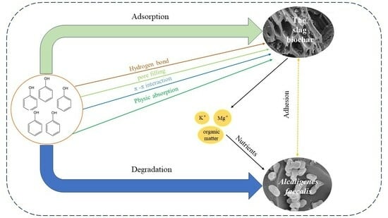
Phenol is widely utilized in various industries such as pharmaceuticals, textiles, coal gasification, leather production, resin synthesis, pulp and paper manufacturing, paint production, and wood processing [1]. It is important to note that phenol has also been classified as a priority pollutant by the United States Environmental Protection Agency (US EPA) [2]. Phenol has a wide range of toxic effects for both acute and chronic exposures, is carcinogenic, and is one of the endocrine-disrupting compounds that cause serious hazardous damage to human health and the environment [3,4].
At present, the primary methods employed for treating phenol-containing wastewater encompass adsorption, biodegradation, solvent extraction, and chemical oxidation [5]. Among these methods, biodegradation has emerged as the most promising, practical, and cost-effective approach for effectively removing phenol from chemical production processes [6]. However, the application of bioremediation at contaminated sites is greatly limited because microorganisms usually exhibit unsatisfactory survivability and activity in the face of load shock and highly heterogeneous environmental media [7]. To tackle these concerns, one potential solution is the utilization of cell immobilization in microbial treatment, where microorganisms are confined to a specific spatial region. This confinement ensures the sustained biological activity and rapid proliferation rates, thereby enhancing the overall efficiency [8].
Biochar, a carbon-rich and porous substance, is created through the controlled thermal conversion of biomass in a low-oxygen environment, typically below temperatures of 600 °C [9]. Due to its large specific surface area and the presence of negatively charged organic functional groups, it possesses strong adsorption capacity, which makes it an effective adsorbent for the removal of various pollutants from wastewater, including metals and organic compounds [10]. It is highly efficient, low cost, easy to apply, and produces few byproducts compared to conventional alternatives [11]. However, biochar obtained by the high-temperature cracking of biomass has limited functional groups and porosity, so it is modified before and after biomass cracking by oxidation, sulfonation, amination, and compounding to adjust the surface functionality and introduce abundant functional groups or nanomaterials into the surface of biochar to change its porosity and surface functionality [12,13].
Immobilized microbial technology is a bioengineering technique that uses physical or chemical means to confine free microorganisms to a specific area so that they can maintain a relatively high level of biological activity in a highly dense state and can be used repeatedly. The most common microorganisms immobilized by carriers are fungi as well as bacteria [14]. Research has shown that the biomass of calcium alginate-immobilized Bacillus cereus strain, capable of degrading phenolic compounds in petroleum wastewater, exhibits excellent biodegradation efficiency. Microbial treatment has been found to reduce Chemical Oxygen Demand (COD) levels and phenolic compound concentrations by a remarkable 95% [15]. Although traditional immobilization techniques have their own advantages, they all have shortcomings. Therefore, to improve the immobilization effect and treatment efficiency, compound immobilization methods have been derived from basic immobilization methods, which can enhance immobilization techniques by co-fixing mixed strains of bacteria, or bacteria and algae, or by improving the carriers of immobilized microorganisms.
Biochar is an ideal immobilization carrier, characterized by its large surface area, porous structure, and strong stability [16]. The immobilization of microorganisms with biochar occurs through the coordinated impregnation of pollutant-degrading microorganisms and biochar. This synergistic approach enhances the mass transfer of pollutants from the contaminated environment to the degrading microbial community, while promoting the enrichment of degrading bacteria to form a biofilm [17]. Zhuang [18] investigated Fe3O4 nanoparticles loaded with bamboo charcoal (Fe3O4/BC) and immobilized Streptomyces sp. N01 for quinoline removal from water. The results showed that bamboo charcoal enhanced the enzymatic activity as a barrier for bacteria, while Fe3O4 nanoparticles improved the cell permeability; thus, the quinoline degradation efficiency was significantly improved. However, there is still a scarcity of systematic studies on the removal of phenol by microorganisms immobilized on modified biochar. Singh and Balomajumder [19] investigated the effect of the simultaneous removal of S. odorifera (MTCC 5700) immobilized on the surface of coconut shell-activated carbon (CSAC) from single- and two-component aqueous solutions in an intermittent reactor on phenol. The simultaneous removal of phenol using S. odorifera (MTCC 5700) immobilized on the CSAC biosorbent surface exhibited superior performance in the binary component system compared to the single component biosorbent system.
In this paper, Andrographis herb residue, which is not easily disposed, was selected as the raw material for biochar. Biochar pyrolyzed under different temperatures and modified with Fe3O4 was selected for the adsorption and immobilization of bacteria to determine the differences caused by these factors and their effects on bacterial immobilization. Simultaneous degradation of phenol by dioxygenases requires a metal cofactor, most commonly Fe(II) or Fe(III) [20]. The nanomaterial Fe3O4 was chosen for modification to form loaded biochar. The biochar was used as a carrier to adsorb the immobilized strain JH1 to investigate the mechanisms of phenol removal and to analyze the performance of multiple recycling.
2. Materials and Methods
2.1. Chemicals and Culture Medium
Phenol (>99% purity) was acquired from Aladdin (Shanghai, China). All other chemicals used in this study were of analytical grade and were obtained from local suppliers. The Luria-Bertani (LB) medium consisted of 10.0 g/L tryptone, 5.0 g/L yeast extract, and 10.0 g/L NaCl (pH = 7.0). The mineral salt medium (MSM) was comprised of 1.0 g/L Na2HPO4, 0.5 g/L KH2PO4, 0.03 g/L MgSO4·7H2O, and 1 mL of trace element solution (pH = 7.0). The trace element solution contained 0.2 g/L MnCI2·4H2O, 0.24 g/L CoCl2·6H2O, 0.15 g/L NaMoO4·2H2O, 0.15 g/L FeCl3·6H2O, 0.16 g/L ZnSO4·7H2O, and 0.2 g/L NiCl2·6H2O. Addition of AGAR (15 g/L) to form solid medium. The LB medium or MSM medium were incubated in an orbital shaker at 30 °C and 150 rpm.
A dominant phenol-degrading strain was domesticated from coking wastewater in Nanchang, Jiangxi Province, China, and named Alcaligenes faecalis strain JH1. In brief, 30 mL of coking wastewater sludge supernatant was added to a conical flask containing 70 mL of MSM medium with 10 mg phenol. The samples were then incubated in an orbital shaker at 30 °C and 150 rpm. After 2 to 7 days of culturing, a 30 mL portion of the culture solution was transferred and underwent subculturing under the same conditions. After subculturing for 6 generations, strain JH1 could metabolize and remove phenol effectively with phenol as the only carbon source.
2.2. Fe3O4 Nanoparticle-Modified Biochar Preparation
Andrographis paniculata slag residue was obtained from the Guangdong Pharmaceutical University laboratory. The biomass materials were prepared by the slow pyrolysis method. First, the dried biomass materials were processed by crushing them using a high-speed multifunctional crusher, sifted through a 2 mm sieve, and then transferred to a quartz-tube furnace (SK-G06123K, Tianjin, China). Then, under a constant supply of nitrogen, biomass materials were pyrolyzed separately at 300 °C, 500 °C and 700 °C for 2 h at a heating rate of 5°/min. After cooling to room temperature, they were ground through a 100-mesh sieve, washed with deionized water, and dried for 12 h in an oven at 80 °C. Finally, the biochars of medicinal residue (Y3, Y5, Y7) were packed in Ziplock bags and stored in a desiccator.
In this study, Y7 was modified by Fe3O4 nanoparticles [1]: 5 g biochars were suspended in a mixture of 100 mL FeCl3·6H2O (2.9 g, 10.8 mmol) and FeSO4·7H2O (6.0 g, 21.5 mmol) at 70 °C, and then 10 mL NaOH solution (5 mol/L) was added to precipitate Fe3O4 nanoparticles under an N2 atmosphere. The obtained materials were dried for 24 h in an oven at 70 °C and expressed as Fe-Y7. The chemical formula of the reaction process is Fe2+ +Fe3+ + 8OH− = Fe3O4↓ + 4H2O.
2.3. Material Characterization
The percentage of C, H, N, and S in the elemental composition of biochar was directly determined by a UNICUBE elemental analyzer, and the content of O could be calculated by difference subtraction. The specific surface area and pore size of biochar were analyzed using an automatic specific surface and porosity analyzer by measuring the adsorption/desorption isotherm of N2 at 77 K. Zeta (ζ) potential measurements were carried out using a NanoBrook Omni instrument (Bruker, Billerica, MA, USA). The surface of the sample was analyzed using the Brunauer–Emmett–Teller (BET) method with N2 gas adsorption at 77.3 K, utilizing the high-speed surface analyzer ASAP 2460 (Micromeritics, Mack Instruments Ltd., Norcross, GA, USA). X-ray diffraction (XRD) patterns were acquired using an X-ray diffractometer (Empyrean, PANalytical, Almelo, The Netherlands) at a scanning range of 2θ from 10° to 80°, and a scanning rate of 10°/min. These measurements were performed to analyze the crystal structure of the biochar. X-ray photoelectron spectroscopy (XPS) using the K-Alpha instrument (Thermo Scientific, Waltham, MA, USA) was employed to analyze the surface chemical composition of the modified biochar as well as the chemical status of iron. The obtained data were analyzed and processed using Advantage 5.5 software. The morphology was observed by transmission electron microscopy (JEM 2100, JEOL, Tokyo, Japan) at various magnifications with an accelerating voltage of 5.0 kV. The obtained images were then used to determine the formation of Fe3O4 nanoparticles on the modified biochar by Digital Micrograph software(Digital Micrograph 2021).
2.4. Immobilization of Strain JH1 to Biochar
Strain JH1 was cultured in LB medium and incubated on a shaking table for 24 h at 30 °C and 150 rpm. The bacteria were centrifuged at 5000 r/min, cleaned with PBS, collected, and suspended again to achieve the logarithmic growth phase (1.2 × 10⁹ CFU/mL). The biochar and cell suspension of strain JH1 were mixed at a ratio of 5:100 (w/v) and shaken on a shaking table for 24 h at 30 °C and 150 rpm to make the bacteria adsorb and adhere on the biochar, and then centrifuged at 1000 rpm for 10 min. The supernatant was filtered and dried at 30 °C. These four biochar-immobilized bacteria were named JY3, JY5, JY7, and JFe-Y7.
2.5. Determination of the Microbial Biomass
The amount of immobilized strain JH1 on the biochar was expressed by measuring biological phosphorus content [21]. Biochar (0.5 g) attached to microorganisms was used to extract phospholipids from cell membranes in a separation funnel containing a mixture of chloroform, ethanol, and water with a volume ratio of 1:2:0.8. The extraction time was 2–24 h. Then, by adding more chloroform and water, the mixture was partitioned into a chloroform phase containing lipids and a methanol–water phase, resulting in a final ratio of chloroform–methanol–water of 1:1:0.9. After standing for an additional 12 h, the lipid-containing chloroform was divided into 50 mL colorimetric tubes, and the chloroform was evaporated in 70 °C water baths. Ammonium molybdate spectrophotometry (TU-1901, Beijing General Instrument Co., Ltd., Beijing, China) was used to determine the content of biological phosphorus. The final result was expressed as nmol P/g biochar.
2.6. Biosorption and Reusability Studies: Batch Experiments
The biosorption experiments were conducted in 100 mL Erlenmeyer flasks, where 0.2 g of either biochars or biochar-immobilized cells were added to 20 mL of phenol solutions. Additionally, 5 mL of cell suspension (1.2 × 10⁹ CFU/mL, Dilution plate counting was used to obtain) was incorporated as the background. The mixture was incubated on a shaking table with a constant agitation rate of 150 rpm at 30 °C for 24 h, unless otherwise stated. The impact of several factors, such as initial phenol concentration, pH, and temperature, on the degradation of phenol by immobilized cells was investigated. Briefly, the experimental conditions were as follows: initial phenol concentrations of 300, 400, 500, and 600 mg/L; incubation temperatures of 25, 30, 35, and 40 °C; and initial pH levels of 5.0, 6.0, 7.0, 8.0, and 9.0. Samples were periodically collected and filtered using a 0.22 μm organic filter membrane (PES) for subsequent analysis of residual phenol concentration.
Biochar-immobilized cells were repeatedly used in phenol degradation for 8 cycles to test their reusability. In brief, 0.5 g of biochar-immobilized cells were incubated in 20 mL MSM medium containing 300 mg/L phenol at pH 7.0 on a shaking table at 150 rpm at 30 °C in 100 mL Erlenmeyer flasks. Samples were collected periodically through a 0.22 μm organic filter membrane for residual phenol concentration analysis. The first cycle ended when phenol reached 99% in solution within 0 to 4 days. The Erlenmeyer flask was allowed to stand for 1–2 min to precipitate the biochar, after which the supernatant was withdrawn with a syringe. Then, sterilized distilled water and phenol were added to adjust the phenol concentration in the solution to 300 mg/L again. Then, the second cycle was started, and the experimental operation was the same as the first cycle, repeated for eight cycles. The eight cycles were named 1C, 2C, 3C, 4C, 5C, 6C, 7C, and 8C. In particular, during the experiment at 8 °C, samples were taken every 2 h to determine the concentration of phenol in the solution, and the sampling was stopped when the phenol concentration of all solutions dropped to 0. At the same time, different cycles were selected for scanning electron microscope (SEM) imaging and to determine the biomass of biochar-immobilized cells.
2.7. Effect of Biochar Water-Extractable Organic Carbon on Strain JH1
To study whether the water-extractable organic carbon (WEOC) of biochar affects the degradation of bacteria, the WEOC of biochar was investigated according to the study of Graber [22]. Biochar was added to the MSM medium at a ratio of 0.1% (w/v), and then the medium was oscillated at 150 rpm at 30 °C for 24 h. The biochar was then removed with a 0.22 μm organic filter membrane to obtain a filtrate containing biochar WEOC and MSM medium. A 6 × 10⁷ CFU/mL cell suspension was added to 50 mL of the above filtrate containing 300 mg/L phenol, and MSM medium without cells was used for the control experiment. All media were incubated in the dark for 24 h at 30 °C, 150 rpm, pH 7 on a shaking table. Samples were collected periodically through a 0.22 μm organic filter membrane for residual phenol concentration analysis. Meanwhile, after 24 h of degradation, the enzyme activities of bacteria in the solution were determined. Catechol 2,3-dioxygenase and catechol 1,2-dioxygenase activities were determined by measuring the production of either 2-hydroxymuconic semialdehyde at 375 nm or muconic acid at 260 nm [23] using a UV–VIS spectrophotometer (TU-1901, PERSEE, China).
2.8. Fourier Transform Infrared Spectroscopy (FTIR) Analysis
Fourier transform infrared spectroscopy (FTIR, Nicolet is 50, Thermo Scientific, Waltham, MA, USA) was used to detect whether extracellular polymeric substances (EPS) were generated during the interaction between bacteria and biochar. Alternatively, it can be used to determine the group types of biochar before and after the adsorption of phenol to measure the mechanism of biochar adsorption of phenol. All FTIR spectra were collected within the wavenumber range of 400~4000 cm−1. Each spectrum was obtained by averaging 32 scans, and the spectral resolution was set at 4 cm−1. The spectra were displayed in terms of absorbance.
2.9. Scanning Electron Microscopy (SEM) Analysis
The morphology of biochar-immobilized bacteria was observed by scanning electron microscopy (Merlin, Zeiss, Jena, Germany). The biochar-immobilized bacteria were fixed in 2.5% (w/v) glutaraldehyde phosphate buffer at 4 °C for 12 h, and then the residual glutaraldehyde was washed twice with PBS. Then, the dehydrated samples were dehydrated with a multiconcentration gradient (30%, 50%, 70%, 90%, and 100%) of ethanol and incubated for 10~15 min at each stage. The dehydrated samples were freeze-dried in a vacuum. The distribution was observed by a scanning electron microscope after spraying gold.
2.10. Analytical Methods of Phenol
The concentration of phenol was determined using the 4-AAP spectrophotometric method with a UV–VIS spectrophotometer (TU-1901, PERSEE, China) at 510 nm [24]. Samples were centrifuged at 6000 rpm for 10 min, and the supernatant was collected to analyze the phenol concentration. Biochar-containing samples were filtered through a 0.22 μm organic filter membrane, and the filtrate was collected to analyze the phenol concentration. Microsoft Excel 2019 and Origin 2017 were used for data processing and charting, respectively. pH was measured using a pH meter (ST300, OHAUS, USA).
3. Results and Discussion
3.1. Characterization of Biochar and the Modified Biochar
Table 1 shows the yield, elemental composition, and elemental content ratio of the Andrographis paniculata medicinal residue biochar prepared at different pyrolysis temperatures. The biochar yield of the medicinal residue decreases with increasing charring temperature, mainly because during the pyrolysis process, the biomass decomposes rapidly into volatiles and biochar [12], resulting in the content of volatiles decreasing, while after the charring temperature is >700 °C, the content of volatiles remains approximately the same, while the biochar yield also remains the same [25,26]. The order of strength for stability (O/C) was Fe-Y7 > Y3 > Y7 > Y5, for aromaticity (H/C) was Y7 > Fe-Y7 > Y5 > Y3, and for polarity ((O + N)/C) was Fe-Y7 > Y3 > Y7 > Y5. The aromaticity of Andrographis paniculata medicinal residue biochar decreased with increasing pyrolysis temperature. After biochar modification, Fe-Y7 had stronger stability and polarity, but the aromaticity decreased, which indicated that the decrease in polar functional groups on the surface led to a decrease in polarity.
Analysis of the data presented in Table 2 reveals a clear trend: as the pyrolysis temperature of the Andrographis paniculata medicinal residue biochar increased, both the specific surface area and pore capacity exhibited a notable increase. Additionally, it was observed that the average pore size decreased as the pyrolysis temperature increased. The average pore sizes of Y3 and Y5 ranged from 2 to 50 nm, indicating that their pore sizes were mainly mesopores, while the average pore size of Y7 was close to that of micropores, which was 2.0441 nm. The results indicated that as the pyrolysis temperature increased, the biochar formed more micropores, which effectively increased the specific surface area and contact with contaminants, as well as providing more attachment and proliferation sites for immobilized bacteria [25]. Fe-Y7 has a larger specific surface area and higher pore capacity than Y7, suggesting that the loading of Fe3O4 nanoparticles on the biochar surface increases the contact area with contaminants, resulting in an increase in the overall specific surface area. It also resulted in a larger average pore size and the formation of more mesopores and macropores [27]. The relatively high specific surface area values for Y7 and Fe-Y7, compared to Y3 and Y5, illustrate the effect of temperature on carbonization. In general, the increase in specific surface area values at high-pyrolysis temperatures is mainly due to the removal of volatiles. During pyrolysis, the rapid release of volatiles opens and connects blind and closed pores, as well as forms new cracks, micropores, and mesopores, leading to a significant increase in specific surface area [28].
Figure 1 shows the N2 adsorption–desorption isotherm curves of the four biochars. The N2 adsorption–desorption isotherms of the four biochars are all type IV, and the adsorption hysteresis loop appears in the middle section. According to the IUPAC classification, the hysteresis loops of Y7 and Fe-Y7 are of type H4. The hysteresis loops of type H4 have no obvious saturation adsorption plateau, and the N2 adsorption rises rapidly in the low-pressure region (relative pressure P/P0 < 0.1), which is caused by the filling adsorption of micropores, indicating that the biochar contains abundant micropores, narrow fissure pores or laminar structure, and this type of hysteresis loop indicates that the pore structure of biochar is very irregular, mainly consisting of mesopores and macropores.
The zeta potential of the four biochars was measured under different pH conditions, and the pH value corresponding to a zeta potential of 0 was the zero point charge (pHPZC) of the biochar. Figure 2a shows that the isoelectric point of the biochar is lower (pHPZC < 4) [29]. The pHPZC of all four biochars is less than 7.0 and negatively charged, so the surfaces of both phenol and biochar are negatively charged, and electrostatic repulsion is easily generated between them, resulting in biochar maintaining a low adsorption capacity for phenol. However, the good adsorption of phenol by biochar prepared under high-temperature conditions implies that electrostatic adsorption does not play a major role in phenol adsorption by biochar and may also be influenced by strong hydrogen bonds and π–π interactions [30]. For the modified biochar, the pHPZC of Fe-Y7 was higher than that of Y7 by 2.53 and 2.19, respectively.
The crystalline minerals of biochar are mainly SiO2 and CaCO3 at different pyrolysis temperatures Figure 2b. Biochar prepared under low-temperature pyrolysis (Y3) has no obvious crystalline peaks and shows only a small amount of SiO2 crystalline minerals, among which Y3 shows several significant peaks at 2θ values of 15°~25°, which are mainly amorphous C peaks related to the change in crystalline structure of cellulose in biomass [25]. As the pyrolysis temperature increased, the crystalline structure of the drug residue biochar improved, and the content of crystalline minerals increased. The most abundant crystalline mineral in the residue biochar is CaCO3, and there is a most obvious CaCO3 signal peak at a 2θ value of 29.4° (d = 3.03 Å), and the signal peak becomes increasingly intense with increasing temperature. The diffraction peak of SiO2 was enhanced and then weakened with increasing temperature. Compared with the unmodified biochar, the basic crystal structure of the modified biochar is the same, except that the diffraction peaks of Fe3O4 with different crystallographic planes appear, and the 2θ values of Fe-Y7 are 35.5° (d = 2.52 Å) and 47.3° (d = 1.92 Å), which indicates that the biochar is successfully loaded with Fe3O4 nanomaterials.
To further verify the successful loading of Fe3O4 nanomaterials, TEM and XPS analyses were performed. The XPS spectrum of Fe-Y7 is shown in Figure 2c, which shows the presence of Fe 2p (711.74 eV), O 1s (532.88 eV), C ls (285.27 eV) and Si (102.71 eV) on the surface of Fe-Y7. The peaks at 711.06 and 720.95 eV belong to Fe 2p3/2 and Fe 2p1/2, respectively, and are decomposed into four peaks at 711.1, 714.1, 723.7 and 725.3 eV [31], where 714.1 and 723.7 eV are associated with Fe(II) of Fe3O4 and 711.1 and 725.3 eV are associated with Fe(III). It was demonstrated that Fe3O4 nanoparticles containing both Fe(II) and Fe(III) were successfully loaded on the modified biochar surface. TEM (Figure 2d–f) revealed (110) lattice planes of SiO2 with a characteristic spacing of 0.2465 nm, (012) and (104) lattice planes of CaCO3 with a characteristic spacing of 0.39 and 0.30 nm, and (311) and (222) lattice planes of Fe3O4 with characteristic spacings of 0.25 and 0.24 nm. These data correspond to the diffraction peaks of SiO2, CaCO3, and Fe3O4 in the XRD spectra, which also confirm the formation of Fe3O4.
3.2. Fourier Infrared Spectroscopy (FTIR) Analysis of Biochar Adsorption Strain JH1
Once attached to the carrier, bacteria begin to secrete a surrounding matrix called the extracellular polymer (EPS), which is a protective matrix of some polymers, such as polysaccharides, proteins, and nucleic acids, among other polymers. By the FTIR technique, it is possible to detect whether bacteria adhere to the surface of biochar through EPS substances [32]. Cells composed of a large number of proteins contribute to hydrophobicity, while polysaccharides on the cell surface confer hydrophilicity [33]. Figure 3 shows the FTIR plots of biochar before and after the adsorption of A. faecalis JH1. For the mixed region of proteins and fatty acids from 1500~1200 cm−1, there is a significant difference between the residue biochar before and after the adsorption of A. faecalis JH1. The region 1200~900 cm−1 belong to the polysaccharides within the cell wall, and there are significant stretching vibrations of both the residue biochar and the modified Fe-Y7. Thus, FTIR analysis showed that A. faecalis JH1 could adhere to the surface of biochar through substances such as secreted polysaccharides or proteins. Proteins and polysaccharides in EPS play a key role in promoting initial adhesion and biofilm development [34].
The degree of hydrophobicity acquired by bacteria on the substrate affects the number of cells attached to the biochar, while the hydrophilicity/hydrophobicity of the biochar surface also affects the ability of bacteria to adhere; for example, the presence of -CH2 fatty acids on the bacterial surface causes bacteria to selectively adhere to the more hydrophobic biochar [32]. From the (O + N)/C in Table 1, it is clear that Fe-Y7 is more hydrophilic than Y7, which also corresponds to the functional groups on FTIR. Thus, the surface of the pharmaceutical residue biochar has many hydrophobic and hydrophilic groups, and their adsorption of bacteria is a common effect of bacterial extracellular proteins and polysaccharides, such as C-O, -COO-, =C = O, and aromatic Ar-H functional groups [34]. Moreover, Fe-Y7 has a higher adsorption capacity for bacteria than Y7.
3.3. Biomass Fixed by the Adsorption of Biochar
Figure 4 shows the biomass of different biochar adsorption immobilized A. faecalis JH1. From the data in the figure, it can be seen that the amount of bacteria immobilized by Fe-Y7 is the highest with 372.45 nmol P/g biochar, which confirms what was found by FTIR. The number of bacteria immobilized by adsorption was higher for Y5 than for Y3 and Y7 because Y3 and Y7 have more and stronger absorption peaks in the region of 1500–1200 cm−1 in FTIR, with abundant hydrophobic functional groups and therefore strong hydrophobicity, which is not favorable for their adsorption and immobilization of bacteria in the aqueous environment. Therefore, the hydrophilicity and hydrophobicity of biochar affect the amount of fixed biomass.
3.4. Effect of Water-Soluble Organic Carbon from Biochar
To better illustrate the process of phenol removal by biochar-immobilized bacteria, the effect of the water-soluble organic carbon (WEOC) of biochar on the bacterial degradation of phenol was investigated. Figure 5 shows the appearance of water-soluble organic carbon solutions of different biochars, which are colorless and transparent except for Y3, which is light yellow, with a darker color implying more dissolved organic carbon [35]. Generally, the absorbance values of biochar WEOC decreased significantly with increasing biochar pyrolysis temperature [36]. Figure 6a shows the effect of the WEOC of biochar on the bacterial degradation of phenol. As shown by the data at 12 h in the figure, the WEOC of biochar at different temperatures has different effects on the removal of phenol. Compared with the control experiment, WEOC in Y3 and Y7 had a facilitative effect on phenol removal. The water-soluble organic carbon of low-temperature biochar has greater DOC and abundant low molecular weight molecules [37], and these stimulate microbial activity and increase microbial abundance [36], thereby accelerating the degradation of phenol.
After the phenol in solution was degraded within 24 h, the enzymatic activity of the bacteria in solution was measured, as shown in Figure 6b. The results indicate that the pathway of phenol degradation by A. faecalis JH1 relies mainly on catechol 1,2-dioxygenase (C12O) for the production of cis–cis-mucofuranic acid after the ring-opening of catechol [38], and only a small amount of phenol is assimilated by catechol 2,3-dioxygenase (C23O) for the production of 2-hydroxymucofuranic acid semialdehyde. In addition, a comparison with the control bacterial solution revealed that the water-soluble organic carbon of all four biochars promoted the enzymatic activity of the bacteria. The results of Y3 and Y7 were consistent with the data at 12 h in Figure 6a. In contrast, Y5, which had an inhibitory effect on phenol degradation at 12 h in Figure 6b, had stronger enzyme activity measured after 24 h than that of the bacterial solution. This indicates that Y5 is susceptible to the effects of PAHs on water-soluble organic carbon in the short term. However, for Y5 and Fe-Y7, the water-soluble organic carbon under long-term use had a beneficial effect on the enzymatic activity of the bacteria.
3.5. Adsorption and Degradation Kinetics of Phenol
Figure 7 shows the adsorption degradation kinetic curves of different biochar-immobilized A. faecalis JH1. Comparing with the control biochar and bacterial suspension, the biochar-immobilized bacteria showed higher removal of phenol from the solution in different time periods, and even around 96 h, all biochar-immobilized bacterial strains JH1 were able to achieve around 99% degradation of phenol from the solution. However, the control biochar basically reached adsorption saturation at around 48 h, and only a small amount of phenol adsorption would be added with the extension of time, whereas the suspension maintained a low phenol degradation rate all the time, with only 23.4% phenol degradation at 96 h. The control biochar was also found to be a good choice for the adsorption of phenol in the suspension. Therefore, these results suggest that the removal of phenol by biochar-immobilized A. faecalis JH1 is a synergistic effect of adsorption and bacterial degradation, and that biochar itself can adsorb a large amount of phenol, and therefore increase the chance of bacterial contact with phenol and promote bacterial degradation of phenol. In addition, the large number of pores in the biochar provides an important habitat for microorganisms [26] and provides them with shelter, which reduces the toxicity of phenol to microorganisms, and at the same time, the biochar can stimulate the growth and activity of microorganisms, and microorganisms can utilize the ash of the biochar to provide mineral nutrients [28].
3.6. Effect of Temperature, pH, Initial Phenol Concentration, and the Salinity
Figure 8a–d show the effects of temperature, pH, initial concentration, and salinity of phenol on phenol removal by biochar-immobilized strain JH1 for 48 h, respectively. It can be seen from Figure 8 that JY3 shows an extremely high advantage in the removal of phenol, but the high temperature and salinity affect its removal rate of phenol. This result can indicate that the degradation of phenol by JY3 relies mainly on the degradation of suspended bacteria in solution, which is consistent with the results in WEOC. JY5 showed better phenol removal than Y5 at low temperatures, alkalinity, low phenol concentration, and low salinity, suggesting that the removal of phenol by JY5 is susceptible to environmental influences. The lower removal of phenol by JY5 under alkaline conditions may be due to the higher alkalinity of the biochar as the temperature increases [39]. Unlike JY5, JY7 and JFe-Y7 were not greatly affected by the environment, and both showed very high phenol removal rates in different environments, with bacteria still growing and remaining active in the porous structure [40]. Therefore, compared with suspended bacteria, biochar-immobilized bacteria can improve their tolerance in different environments, and they can still grow and maintain strong activity within 48 h at temperatures of 25 °C to 40 °C, pH 5~9, initial phenol concentrations of 300–500 mg/L and salinity of 3%.
3.7. Effect of Recycling Biochar-Fixed Bacteria on Phenol Adsorption
To investigate the reusability of the biochar-immobilized A. faecalis JH1 to confirm its potential for practical applications, the biochar-immobilized bacteria were subjected to recycling experiments. The eight cycles were 96, 60, 48, 48, 36, 48, 24, and 12 h. Seven of these cycles are shown in Figure 8. In the first cycle (1C), the removal of phenol relied mainly on the adsorption of biochar and the bacteria in the solution. As the number of cycles increases, the bacteria use the phenol adsorbed on the biochar as well as nutrients from the biochar itself (e.g., water-soluble organic carbon) to multiply and grow rapidly and adhere to the biochar, and more and more bacteria are adsorbed on the biochar, so that the time spent for the removal of phenol from solution by biochar immobilized bacteria will become shorter and shorter, and thus the long-term phenol removal effect depends more on the biodegradation on the biochar. After the sixth cycle, all biochar-immobilized bacteria could remove 300 mg/L phenol solution within 12 h (Figure 9).
Fe-Y7 showed the most stable removal of phenol and the fastest removal rate (Figure 10). In different cycles, JY3 and JY5 removed phenol from the solution at longer and shorter times, and the removal ability was unstable. In these seven cycles, all biochar-immobilized bacteria could remove phenol from the solution, which may be because biochar as a porous carrier provided enough space for bacterial growth and promoted the growth and activity of bacteria. These results suggest that biochar carrier-immobilized JH1 has great potential for phenol removal, is reusable, and exhibits good stability and durability, in agreement with the results of most recycling studies [18,40].
In contrast, in the 8C cycle (Figure 11), phenol removal by JFe-Y7 was relatively rapid. The dioxygenase of A. faecalis JH1 for phenol degradation uses oxygen as an oxidant to bind two oxygen atoms to the product, while most dioxygenases require a metal cofactor, most commonly Fe(II) or Fe(III) [41]. The addition of appropriate Fe3O4 nanoparticles improves their enzymatic activity [42], small amounts of Fe ions are gradually released to the environment through Fe3O4 nanoparticles, and Fe2+/Fe3+ in solution couples with bacterial oxidation/reduction reactions to accelerate the electron transfer rate [43,44]. Thus, the results demonstrate that Fe3O4 nanoparticle modification accelerates phenol removal as a result of improving the adsorption capacity of phenol or promoting bacterial growth, metabolism, and enzymatic activity.
By performing further SEM analysis, Figure 12a–h shows the 2 k× and 20 k× multiples of 0C, 1C, 4C, and 8C in the cycle, respectively, and the main distribution area of A. faecalis JH1 is on the surface of the biochar and in the internal channels of the pores. At 0C, A. faecalis JH1 adhered to the biochar in relatively scattered and small numbers (Figure 12a,b). However, after one cycle, the bacteria adhering to the biochar surface started to grow slowly using the phenol adsorbed by the biochar and the water-soluble organic carbon of the biochar, splitting and multiplying around it as a single bacterium to become a multicolony, at which time there was no large number of bacteria present on the biochar surface (Figure 12c,d). However, after four cycles, a large number of bacteria were evident on the surface and in the pores of the biochar, and the bacteria multiplied and adhered to the biochar rapidly (Figure 12e,f). With repeated phenol replacement and bacterial growth, a thick and sticky biofilm was established on the biochar surface in the eighth cycle (Figure 12g,h), [42]. The biofilm thickness was in the range of 10~20 µm, and both phenol and oxygen could diffuse into the bacteria for metabolic activities [45]. The SEM images of these four biochar-immobilized A. faecalis JH1 were generally consistent, and the growth and adhesion of bacteria on biochar showed a similar variation pattern. Thus, biochar is an ideal carrier with a large specific surface area for bacterial adhesion and growth, is nontoxic and cost-effective, and additionally facilitates oxygen and substrate delivery through immobilized cells and enhances the activity of immobilized bacteria.
4. Conclusions
The microstructure of Andrographis paniculata medicinal residue is a honeycomb with a porous structure and a large surface area. Modification by Fe3O4 nanoparticles increases the specific surface area during pyrolysis, and the data demonstrate the successful loading of Fe3O4 nanoparticles onto the surface of Fe-Y7. The adsorption of all four biochars on E. Alcaligenes faecalis strain JH1 relies on the combined action of bacterial extracellular proteins and polysaccharides, and Fe-Y has a higher adsorption capacity for bacteria. The biochar-immobilized bacteria were more tolerant than the suspended bacteria in different environments. During the recycling process, with the increase in the number of cycles, the bacteria will form biofilms on the biochar, and the formed biofilms make the biochar-immobilized bacteria still have a certain removal effect on phenol after several cycles, among which the removal effect of Jeffe-Y7 is relatively better. The results showed that Fe3O4 nanoparticle-modified biochar-immobilized microorganisms could promote the degradation of phenol with good reproducibility, and therefore, this technology has potential application in the treatment of phenol-containing wastewater. It can be used in industry to recycle phenol wastewater, which is less expensive, saves treatment costs and does not produce other environmental pollution. The removal of phenol by the biochar-immobilized A. faecalis JH1 was a dual action of adsorption-degradation. The biochar mainly relied on π–π interactions and hydrogen bonding to adsorb phenol, which was first adsorbed by the biochar before being metabolically degraded by the bacteria on the biochar.
Author Contributions
Conceptualization, J.X.; Methodology, Z.Z.; Software, Z.Z. and J.X.; Validation, M.L. and J.W.; Formal analysis, Z.Z.; Investigation, Z.Z., J.X., M.L. and J.W.; Data curation, Z.Z.; Writing—original draft, Z.Z.; Writing—review and editing, T.Z.; Supervision, T.Z.; Project administration, T.Z.; Funding acquisition, T.Z. All authors have read and agreed to the published version of the manuscript.
Funding
This study was financially supported by the National Natural Science Foundation of China Projects (U21A2003), and National Key R&D Program of China (2020YFC1808803).
Data Availability Statement
The data that supports the findings of this study are available within the article.
Conflicts of Interest
The authors declare no conflict of interest.
References
- Panigrahy, N.; Priyadarshini, A.; Sahoo, M.M.; Verma, A.K.; Daverey, A.; Sahoo, N.K. A comprehensive review on eco-toxicity and biodegradation of phenolics: Recent progress and future outlook. Environ. Technol. Innov. 2022, 27, 102423. [Google Scholar] [CrossRef]
- Sahoo, S.K.; Das, A.A.; Deka, D.; Naik, B.; Kumar Sahoo, N. Organic-inorganic hybrid hydroquinone bridged v-cds/hap/pd-tcpp: A novel visible light active photocatalyst for phenol degradation. J. Mol. Liq. 2021, 339, 116721. [Google Scholar] [CrossRef]
- Darbre, P.D. The history of endocrine-disrupting chemicals. Endocr. Metab. Res. 2019, 7, 26–33. [Google Scholar] [CrossRef]
- Martínková, L.; Kotik, M.; Marková, E.; Homolka, L. Biodegradation of phenolic compounds by basidiomycota and its phenol oxidases: A review. Chemosphere 2016, 149, 373–382. [Google Scholar] [CrossRef]
- Kujawski, W.; Warszawski, A.; Ratajczak, W.; Porębski, T.; Capała, W.; Ostrowska, I. Removal of phenol from wastewater by different separation techniques. Desalination 2004, 163, 287–296. [Google Scholar] [CrossRef]
- Wu, J.; Yu, H.Q. Biosorption of 2,4-dichlorophenol from aqueous solution by phanerochaete chrysosporium biomass: Isotherms, kinetics and thermodynamics. J. Hazard. Mater. 2006, 137, 498–508. [Google Scholar] [CrossRef]
- Biswas, B.; Sarkar, B.; Rusmin, R.; Naidu, R. Bioremediation of pahs and vocs: Advances in clay mineral–microbial interaction. Environ. Int. 2015, 85, 168–181. [Google Scholar] [CrossRef]
- Karel, S.F.; Libicki, S.B.; Robertson, C.R. The immobilization of whole cells: Engineering principles. Chem. Eng. Sci. 1985, 40, 1321–1354. [Google Scholar] [CrossRef]
- Kim, P.; Johnson, A.M.; Essington, M.E.; Radosevich, M.; Kwon, W.-T.; Lee, S.-H.; Rials, T.G.; Labbé, N. Effect of pH on surface characteristics of switchgrass-derived biochars produced by fast pyrolysis. Chemosphere 2013, 90, 2623–2630. [Google Scholar] [CrossRef]
- Jin, Z.; Xiao, S.; Dong, H.; Xiao, J.; Tian, R.; Chen, J.; Li, Y.; Li, L. Adsorption and catalytic degradation of organic contaminants by biochar: Overlooked role of biochar’s particle size. J. Hazard. Mater. 2022, 422, 126928. [Google Scholar] [CrossRef] [PubMed]
- Beesley, L.; Moreno-Jimenez, E.; Gomez-Eyles, J.L.; Harris, E.; Robinson, B.; Sizmur, T. A review of biochars’ potential role in the remediation, revegetation and restoration of contaminated soils. Environ. Pollut. 2011, 159, 3269–3282. [Google Scholar] [CrossRef]
- Liu, W.J.; Jiang, H.; Yu, H.Q. Development of biochar-based functional materials: Toward a sustainable platform carbon material. Chem. Rev. 2015, 115, 12251. [Google Scholar] [CrossRef]
- Xiao, X.; Chen, B.; Chen, Z.; Zhu, L.; Schnoor, J.L. Insight into multiple and multilevel structures of biochars and their potential environmental applications: A critical review. Environ. Sci. Technol. 2018, 52, 5027–5047. [Google Scholar] [CrossRef] [PubMed]
- Girijan, S.; Kumar, M. Immobilized biomass systems: An approach for trace organics removal from wastewater and environmental remediation—sciencedirect. Curr Opin Environ Sci Health 2019, 12, 18–29. [Google Scholar] [CrossRef]
- Banerjee, A.; Ghoshal, A.K. Biodegradation of real petroleum wastewater by immobilized hyper phenol-tolerant strains of Bacillus cereus in a fluidized bed bioreactor. 3 Biotech 2016, 6, 137. [Google Scholar] [CrossRef]
- Manya, J.J. Pyrolysis for biochar purposes: A review to establish current knowledge gaps and research needs. Environ. Sci. Technol. 2012, 46, 7939. [Google Scholar] [CrossRef]
- Wu, C.; Zhi, D.; Yao, B.; Zhou, Y.; Yang, Y.; Zhou, Y. Immobilization of microbes on biochar for water and soil remediation: A review. Environ. Res. 2022, 212, 113226. [Google Scholar] [CrossRef] [PubMed]
- Zhuang, H.; Han, H.; Xu, P.; Hou, B.; Jia, S.; Wang, D.; Li, K. Biodegradation of quinoline by streptomyces sp N01 immobilized on bamboo carbon supported Fe3O4 nanoparticles. Biochem. Eng. J. 2015, 99, 44–47. [Google Scholar] [CrossRef]
- Singh, N.; Balomajumder, C. Equilibrium isotherm and kinetic studies for the simultaneous removal of phenol and cyanide by use of S. Odorifera (Mtcc 5700) immobilized on coconut shell activated carbon. Appl. Water Sci. 2017, 7, 3241–3255. [Google Scholar] [CrossRef]
- Wu, Z.; Shen, J.; Li, W.; Li, J.; Xia, D.; Xu, D.; Zhang, S.; Zhu, Y. Electron self-sufficient core-shell biocl@fe-biocl nanosheets boosting Fe(III)/Fe(II) recycling and synergetic photocatalysis-fenton for enhanced degradation of phenol. Appl. Catal. B Environ. 2023, 330, 122642. [Google Scholar] [CrossRef]
- Yang, H.J.; Yang, Z.M.; Xu, X.H.; Guo, R.B. Increasing the methane production rate of hydrogenotrophic methanogens using biochar as a biocarrier. Bioresour. Technol. 2020, 302, 122829. [Google Scholar] [CrossRef]
- Graber, E.R.; Tsechansky, L.; Lew, B.; Cohen, E. Reducing capacity of water extracts of biochars and their solubilization of soil Mn and Fe. Eur. J. Soil Sci. 2014, 65, 162–172. [Google Scholar] [CrossRef]
- Liu, Z.; Yang, H.; Huang, Z.; Zhou, P.; Liu, S.-J. Degradation of aniline by newly isolated, extremely aniline-tolerant Delftia sp. AN3. Appl. Microbiol. Biotechnol. 2002, 58, 679–682. [Google Scholar] [CrossRef]
- Yu, J.; Tao, D.; Yu, S.; Kai, Y.; Wang, H. Biodegradation of phenol by entrapped cell of debaryomyces sp. With nano-Fe3O4 under hypersaline conditions. Int. Biodeterior. Biodegrad. 2017, 123, 37–45. [Google Scholar]
- Keiluweit, M.; Nico, P.S.; Johnson, M.G.; Kleber, M. Dynamic molecular structure of plant biomass-derived black carbon (biochar). Environ. Sci. Technol. 2010, 44, 1247–1253. [Google Scholar] [CrossRef]
- Ahmad, M.; Ok, Y.S.; Rajapaksha, A.U.; Lim, J.E.; Kim, B.Y.; Ahn, J.H.; Lee, Y.H.; Al-Wabel, M.I.; Lee, S.-E.; Lee, S.S. Lead and copper immobilization in shooting range soil using soybean stover-and pine needle-derived biochars: Chemical, microbial and spectroscopic assessments. J. Hazard. Mater. 2016, 301, 179–186. [Google Scholar] [CrossRef]
- Jin, X.; Liu, R.; Wang, H.; Han, L.; Qiu, M.; Hu, B. Functionalized porous nanoscale Fe3O4 particles supported biochar from peanut shell for pb(ii) ions removal from landscape wastewater. Environ. Sci. Pollut. Res. 2022, 29, 37159–37169. [Google Scholar] [CrossRef] [PubMed]
- Liu, L.; He, N.; Borham, A.; Zhang, S.; Xie, R.; Zhao, C.; Hu, J.; Wang, J. The effect of iron-modified biochar on phosphorus adsorption and the prospect of synergistic adsorption between biochar and iron-oxidizing bacteria: A review. Water 2023, 15, 3315. [Google Scholar] [CrossRef]
- Cheng, C.; Lehmann, J.; Engelhard, M.H. Natural oxidation of black carbon in soils: Changes in molecular form and surface charge along a climosequence. Geochim. Cosmochim. Acta 2008, 72, 1598–1610. [Google Scholar] [CrossRef]
- Du, J.; Sun, P.; Feng, Z.; Zhang, X.; Zhao, Y. The biosorption capacity of biochar for 4-bromodiphengl ether: Study of its kinetics, mechanism, and use as a carrier for immobilized bacteria. Environ. Sci. Pollut. Res. 2016, 23, 3770–3780. [Google Scholar] [CrossRef]
- Liu, G.; Feng, K.; Cui, H.; Li, J.; Wang, M. Mof derived in-situ carbon- encapsulated fe3o4@c to mediate polysulfides redox for ultrastable lithium-sulfur batteries. Chem. Eng. J. 2019, 381, 122652. [Google Scholar] [CrossRef]
- Ramos-Escobedo, G.; Pecina-Treviño, E.; Tokunaga, A.B.; Concha-Guerrero, S.; Ramos-Lico, D.; Guerra-Balderrama, R.; Orrantia-Borunda, E. Bio-collector alternative for the recovery of organic matter in flotation processes. Fuel 2016, 176, 165–172. [Google Scholar] [CrossRef]
- Zhou, Y.; Petrova, S.P.; Edgar, K.J. Chemical synthesis of polysaccharide–protein and polysaccharide–peptide conjugates: A review. Carbohydr. Polym. 2021, 274, 118662. [Google Scholar] [CrossRef] [PubMed]
- Yu, Y.; An, Q.; Zhou, Y.; Deng, S.; Miao, Y.; Zhao, B.; Yang, L. Highly synergistic effects on ammonium removal by the co-system of pseudomonas stutzeri xl-2 and modified walnut shell biochar. Bioresour. Technol. 2019, 280, 239–246. [Google Scholar] [CrossRef] [PubMed]
- Xu, J.; Jian, Z.; Wang, Y.; Fang, C.; Hu, Q. Spatial–seasonal characteristics and influencing factors of dissolved organic carbon and chromophoric dissolved organic matter in poyang lake. Environ. Earth Sci. 2023, 82, 44. [Google Scholar] [CrossRef]
- Li, M.; Liu, M.; Joseph, S.; Jiang, C.; Wu, M.; Li, Z. Change in water extractable organic carbon and microbial plfas of biochar during incubation with an acidic paddy soil. Soil Res. 2015, 53, 763–771. [Google Scholar] [CrossRef]
- Bian, R.; Joseph, S.; Shi, W.; Li, L.; Taherymoosavi, S.; Pan, G. Biochar DOM for plant promotion but not residual biochar for metal immobilization depended on pyrolysis temperature. Sci. Total Environ. 2019, 662, 571–580. [Google Scholar] [CrossRef]
- Jiang, Y.; Wen, J.; Bai, J.; Jia, X.; Hu, Z. Biodegradation of phenol at high initial concentration by alcaligenes faecalis. J. Hazard. Mater. 2007, 147, 672–676. [Google Scholar] [CrossRef]
- Fidel, R.B.; Laird, D.A.; Thompson, M.L.; Lawrinenko, M. Characterization and quantification of biochar alkalinity. Chemosphere 2017, 167, 367–373. [Google Scholar] [CrossRef]
- Liu, Y.; Gan, L.; Chen, Z.; Megharaj, M.; Naidu, R. Removal of nitrate using paracoccus sp. Yf1 immobilized on bamboo carbon. J. Hazard. Mater. 2012, 229–230, 419–425. [Google Scholar] [CrossRef]
- Bugg, T.D.H. Dioxygenase enzymes: Catalytic mechanisms and chemical models. Tetrahedron 2003, 59, 7075–7101. [Google Scholar] [CrossRef]
- Zeng, Q.; Xu, J.; Hou, Y.; Li, H.; Du, C.; Jiang, B.; Shi, S. Effect of Fe3O4 nanoparticles exposure on the treatment efficiency of phenol wastewater and community shifts in sbr system. J. Hazard. Mater. 2021, 407, 124828. [Google Scholar] [CrossRef]
- He, S.; Feng, Y.; Ni, J.; Sun, Y.; Xue, L.; Feng, Y.; Yu, Y.; Lin, X.; Yang, L. Different responses of soil microbial metabolic activity to silver and iron oxide nanoparticles. Chemosphere 2016, 147, 195–202. [Google Scholar] [CrossRef] [PubMed]
- He, S.; Zhong, L.; Duan, J.; Feng, Y.; Yang, B.; Yang, L. Bioremediation of wastewater by iron oxide-biochar nanocomposites loaded with photosynthetic bacteria. Front. Microbiol. 2017, 8, 823. [Google Scholar] [CrossRef] [PubMed]
- Wang, X.; Wang, X.; Liu, M.; Bu, Y.; Zhang, J.; Chen, J.; Zhao, J. Adsorption–synergic biodegradation of diesel oil in synthetic seawater by acclimated strains immobilized on multifunctional materials. Mar. Pollut. Bull. 2015, 92, 195–200. [Google Scholar] [CrossRef]
Figure 1.
N2 adsorption−desorption isotherms of biochars.

Figure 2.
(a) Zeta potential of different biochars, (b) XRD patterns of biochars, (c) XPS spectrum of Fe-Y7 of full scan and Fe 2p, and (d–f) TEM and HR-TEM images.
Figure 2.
(a) Zeta potential of different biochars, (b) XRD patterns of biochars, (c) XPS spectrum of Fe-Y7 of full scan and Fe 2p, and (d–f) TEM and HR-TEM images.

Figure 3.
FTIR spectra of biochars before and after adsorption of A. faecalis JH1.

Figure 4.
Biomass of strain JH1 was immobilized by biochar adsorption.

Figure 5.
Appearance of biochars’ WEOC.

Figure 6.
(a) Effect of biochars’ WEOC on the degradation of phenol by A. faecalis JH1; (b) enzyme activity.
Figure 6.
(a) Effect of biochars’ WEOC on the degradation of phenol by A. faecalis JH1; (b) enzyme activity.

Figure 7.
The adsorption and degradation kinetic curve of biochars immobilized A. faecalis JH1.

Figure 8.
Effect on phenol removal from solution for 48 h. (a) temperature; (b) Ph; (c) initial concentration of phenol; (d) salinity.
Figure 8.
Effect on phenol removal from solution for 48 h. (a) temperature; (b) Ph; (c) initial concentration of phenol; (d) salinity.

Figure 9.
Biochar-immobilized bacteria for recycling.

Figure 10.
Time spent in each cycle to completely remove phenol from the solution.

Figure 11.
Phenol removal kinetics curve at 8C.

Figure 12.
SEM images of different cycles. (a,b) 0C; (c,d) 1C; (e,f) 4C; (g,h) 8C.


Table 1.
Chemical composition and pore structure of biochar samples.
| Samples | Yield (%) | Elemental Composition (%) | Atomic Ratio | ||||||
|---|---|---|---|---|---|---|---|---|---|
| C | H | O | N | S | O/C | H/C | (O + N)/C | ||
| Y3 | 44 | 59.44 | 5.19 | 32.70 | 2.54 | 0.13 | 0.550 | 0.087 | 0.593 |
| Y5 | 29.3 | 67.47 | 3.08 | 27.49 | 1.79 | 0.18 | 0.407 | 0.046 | 0.434 |
| Y7 | 25.1 | 67.51 | 1.71 | 29.18 | 1.43 | 0.17 | 0.432 | 0.025 | 0.453 |
| Fe-Y7 | — | 61.2 | 1.70 | 35.51 | 1.34 | 0.26 | 0.580 | 0.028 | 0.602 |
Table 2.
The pore structure parameters of biochars.
| Samples | Specific Surface Area (m2/g) | Pore Capacity (cm3/g) | Average Aperture (nm) |
|---|---|---|---|
| Y3 | 1.8796 | 0.003046 | 6.4819 |
| Y5 | 3.2917 | 0.005034 | 6.1169 |
| Y7 | 138.8126 | 0.070936 | 2.0441 |
| Fe-Y7 | 164.0975 | 0.097943 | 2.3874 |
Disclaimer/Publisher’s Note: The statements, opinions and data contained in all publications are solely those of the individual author(s) and contributor(s) and not of MDPI and/or the editor(s). MDPI and/or the editor(s) disclaim responsibility for any injury to people or property resulting from any ideas, methods, instructions or products referred to in the content. |
© 2023 by the authors. Licensee MDPI, Basel, Switzerland. This article is an open access article distributed under the terms and conditions of the Creative Commons Attribution (CC BY) license (https://creativecommons.org/licenses/by/4.0/).
Share and Cite
MDPI and ACS Style
Zeng, Z.; Xiao, J.; Li, M.; Wu, J.; Zhang, T. Degradation of Phenol by Immobilized Alcaligenes faecalis Strain JH1 in Fe3O4-Modified Biochar from Pharmaceutical Residues. Water 2023, 15, 4084. https://doi.org/10.3390/w15234084
AMA Style
Zeng Z, Xiao J, Li M, Wu J, Zhang T. Degradation of Phenol by Immobilized Alcaligenes faecalis Strain JH1 in Fe3O4-Modified Biochar from Pharmaceutical Residues. Water. 2023; 15(23):4084. https://doi.org/10.3390/w15234084
Chicago/Turabian StyleZeng, Zhi, Jiahui Xiao, Manzhi Li, Jiahui Wu, and Taiping Zhang. 2023. "Degradation of Phenol by Immobilized Alcaligenes faecalis Strain JH1 in Fe3O4-Modified Biochar from Pharmaceutical Residues" Water 15, no. 23: 4084. https://doi.org/10.3390/w15234084
Note that from the first issue of 2016, this journal uses article numbers instead of page numbers. See further details here.